Chapter: The Massage Connection ANATOMY AND PHYSIOLOGY : Skeletal System and Joints
The Thorax
THE THORAX
The bones that form the thoracic cage are the ster-num, ribs, and vertebrae. The thoracic cage protectsthe heart, lungs, and other organs. It provides attach-ment to muscles that stabilize the vertebral column and the pectoral girdle. Muscles that produce respi-ratory movements are also attached. The thorax is narrower superiorly and is flattened anteroposteri-orly. Costal (hyaline) cartilage, present anteriorly, connects the ribs to the sternum.
The Sternum
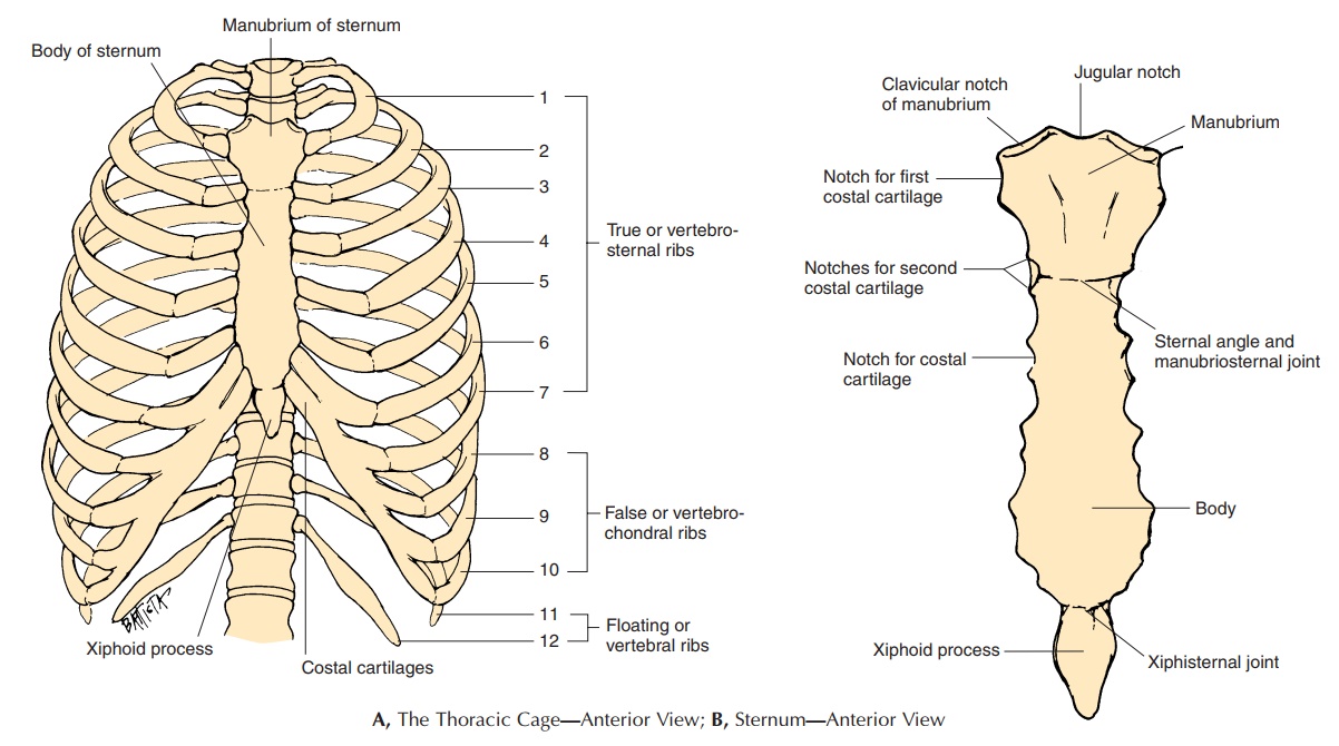
The sternum, or breastbone (see Figures 3.20A and B), lies in the anterior aspect of the thoracic cage, in the midline. The broader, upper part, the manubrium, ar-ticulates with the clavicles and first ribs. A shallow de-pression in its most superior part is the suprasternal, or jugular notch. On the lateral aspect of the manubrium, theclavicular notches are depressions that articulate with the clavicles to form the sterno-clavicular joint. The first two ribs articulate with the manubrium to form the sternocostal joints at the costal notches.
The body (corpus) of the sternum is attached to the inferior surface of the manubrium. A slight eleva-tion that can be felt at this junction is referred to as
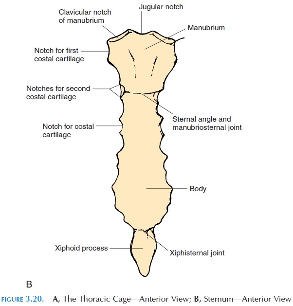
The body articulates with the costal cartilages of ribs 2–7 at the costal notches. The most inferior part of the sternum is the xiphoidprocess, to which the diaphragm and the rectus ab-dominus muscles are attached. The sternal angle is an important landmark because the second costal cartilage is attached to the sternum at this point. The ribs and intercostal spaces below this point can eas-ily be counted from here. It also indicates the loca-tion where the trachea divides into the two primary bronchi. The sounds made by the closing of the aor-tic and pulmonary valves (second heart sound) are best heard in the second intercostal spaces.
The Ribs
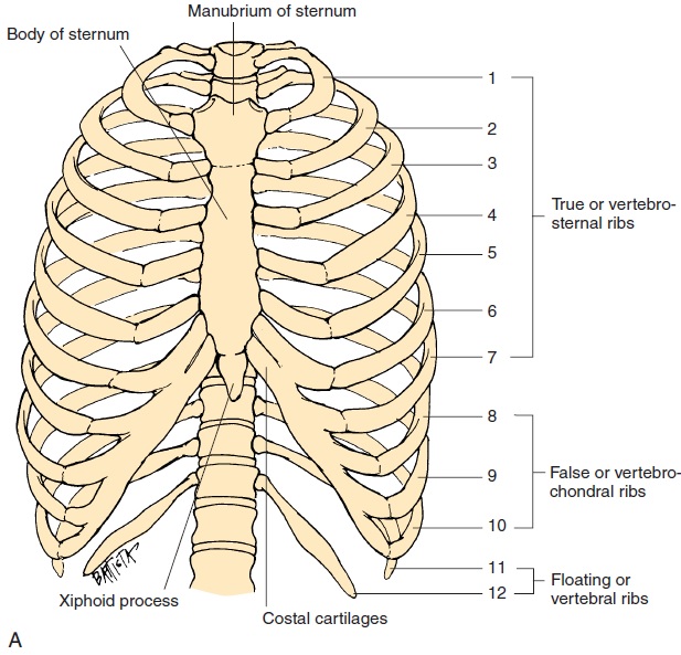
The ribs (Figure 3.20 A and C) are long, flat, and curved. They extend from the thoracic vertebrae to the middle of the thoracic cavity. The ribs are con-nected to the sternum by costal cartilages. The first seven pairs are longer than the others. They are known as true ribs or vertebrosternal ribs because each of these ribs have individual costal cartilages that connect them to the sternum. Ribs 8–12 are known as false ribs or vertebrochondral ribs be-cause they are not connected to the sternum individ-ually, instead the costal cartilages of the ribs 8–10 are fused together before they reach the sternum. Ribs 11 and 12 are not attached to the sternum; they are only attached to the vertebrae. These ribs are called float-ing, or vertebral ribs.
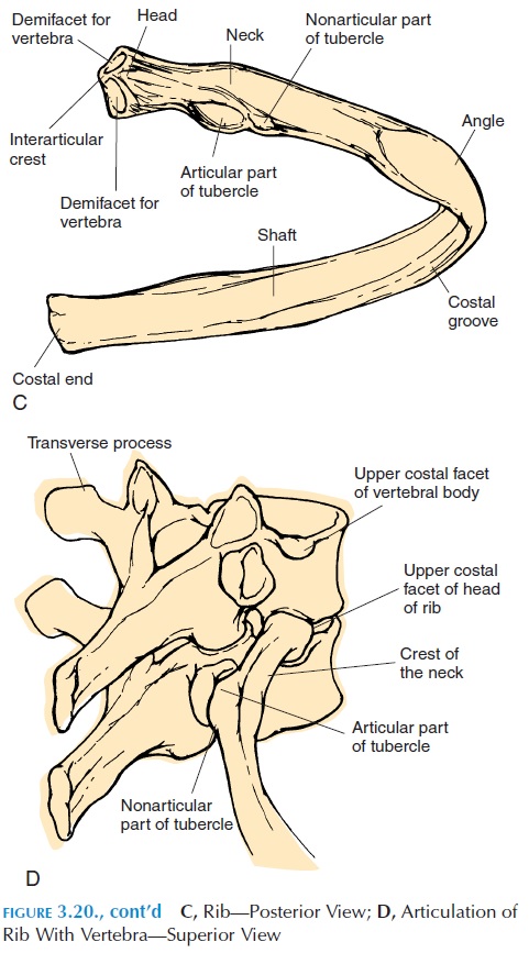
Typically, the posterior end of the rib has a head with two facets that articulate with the facets on the bodies of the vertebrae to form the vertebrocostaljoint. Lateral to the head is the constricted portion called the neck. On the posterior aspect of the neck, there is a short projection, the tubercle. The articu-lar part of the tubercle articulates with the facet on the transverse process of the lower of the two tho-racic vertebrae (to which the head articulates). The neck continues on as the body, or shaft. The shaft curves anteriorly and medially beyond the tubercle, forming the costal angle. The superior and inferior surfaces of the ribs give attachment to the intercostal muscles. The space between any two ribs is known as the intercostal space. The intercostal muscles, blood vessels, and nerves are located here.
There is a groove on the internal aspect of the rib, the costal groove, in which the intercostal nerves and blood vessels lie.
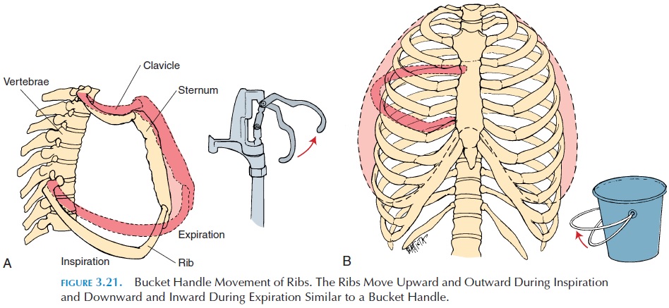
The positioning of the ribs resembles that of a bucket handle (see Figure 3.21). If the ribs are pushed down, the transverse diameter of the thorax is de-creased. It also results in the sternum being pulled in-ward, decreasing the anteroposterior diameter. If the ribs are pulled upward, there is an increase in the transverse and anteroposterior diameter. This is how the thoracic capacity is altered during respiration. The presence of the costal cartilages makes the tho-racic cavity flexible and able to withstand sudden im-pact; however, severe blows can fracture the ribs.
Related Topics