Chapter: The Massage Connection ANATOMY AND PHYSIOLOGY : Skeletal System and Joints
The Hip Joint
THE HIP JOINT
Articulating Surfaces and Type of Joint
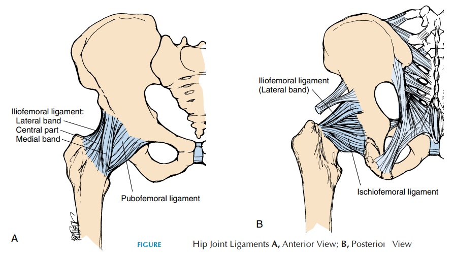
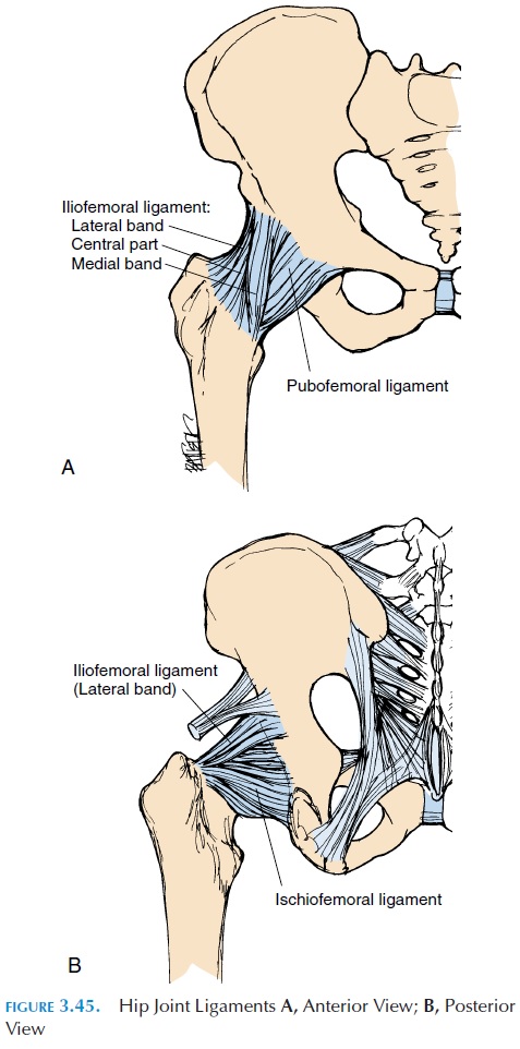
The hip joint, also referred to as the acetabulofemoral or iliofemoral joint, is one of the most stable joints be-cause the articular surfaces of the rounded head of the femur and the acetabulum of the pelvis fit well into each other. The acetabulum is further deepened by the fibrocartilage (acetabular labrum) located in the ac-etabulum. In addition to shape of the articular surface, the hip joint, similar to the shoulder, has supporting ligaments (see Figure 3.45).
Ligaments and Bursa
The thick capsule is reinforced by strong ligaments. The iliofemoral ligament is a thick band that runs between the anterior inferior iliac spine and the in-tertrochanteric line of the femur. This ligament pre-vents excessive internal and external rotation. When standing, this ligament is twisted and pulled taut and results in “locking” of the joint, allowing the person to stand with little muscle action. The pubofemoralligament extends from the pubic portion of the ac-etabular rim to the inferior portion of the neck of the femur. The ischiofemoral ligament runs between the ischial acetabular rim and the superior portion of the femoral neck.
The transverse acetabular liga-ment runs between the gap in the inferior margin ofthe acetabular labrum. Another ligament, the liga-mentum teres, is located inside the joint capsule andruns between the acetabular notch and a small de-pression (fovea capitis) located in the femoral head.
A few bursae surround the hip joint. The il-iopectineal bursa lies on the anterior aspect of thehip joint, deep to the iliopsoas muscle, as it crosses the joint. It may communicate with the joint cavity of the hip joint. The trochanteric bursaelie over the greater trochanter, deep to the gluteus maximus, re-ducing friction between the bone and muscle.
Possible Movements
The hip permits flexion, extension, adduction, abduc-tion, medial rotation, lateral rotation, and some cir-cumduction.
Range of Motion
Abduction, 45–50°
Adduction, 20–30°
External/lateral rotation, 45°
Internal/medial rotation, 35°
Flexion, 135°
Extension, 30°
Muscles
The action of most muscles around the hip can be de-termined from the location. The flexor muscles are located in the anterior quadrant, the extensors in the posterior quadrant, the adductors in the medial, and the abductors in the lateral quadrant.
Muscles that flex the hip:
Primary flexor
Iliopsoas
Secondary flexors
Rectus femoris Sartorius
Muscles that extend the hip:
Primary extensor
Gluteus maximus
Secondary extensor
Hamstrings
Muscles that abduct the hip:
Primary abductor
Gluteus medius
Secondary abductors
Gluteus minimus Tensor fascia lata
Muscles that adduct the hip:
Primary adductor
Adductor longus
Secondary adductors
Adductor brevis Adductor magnus Pectineus Gracilis
Muscles that rotate the hip laterally: Gluteus maximus
Gluteus medius and minimus (posterior fibers) Muscles that rotate the hip medially:
Adductor magnus, longus, brevis
Gluteus medius and minimus (anterior fibers) Iliopsoas
Physical Assessment
Inspection
The gait should be observed as the person enters the room. It is preferable to have the patient’s body ex-posed waist down. When standing normally, the an-terior superior iliac spine should be level with a slight anterior curvature of the lumbar spine. Absence of the lumbar lordosis may indicate spasm of the mus-cles. Weakness of the abdominal muscles may exhibit an abnormally increased lordosis. Look for muscle wasting and body asymmetry.
Palpation
Bony prominences: Various bony prominences can be easily palpated. These are the anterior superior iliac spines, iliac crest, greater trochanter, and pubic tu-bercles anteriorly. Posteriorly, the posterior superior iliac spine and the ischial tuberosity can be palpated.
Other structures: The inguinal ligament, which runs between the anterior superior iliac spine and the pubic tubercle, marks part of the route taken by the male testis as it descends into the scrotum. Bulges in this region may indicate an inguinal hernia. The femoral artery pulsation can be felt just inferior to the inguinal ligament. The femoral vein lies just me-dial to the artery. Tenderness over the sciatic nerve as it emerges from the sacral region can be palpated.
The active and passive range of motion of the hip should be tested as well, together with discrepancies between the two legs.
Related Topics