Chapter: The Massage Connection ANATOMY AND PHYSIOLOGY : Skeletal System and Joints
Bones of the Face
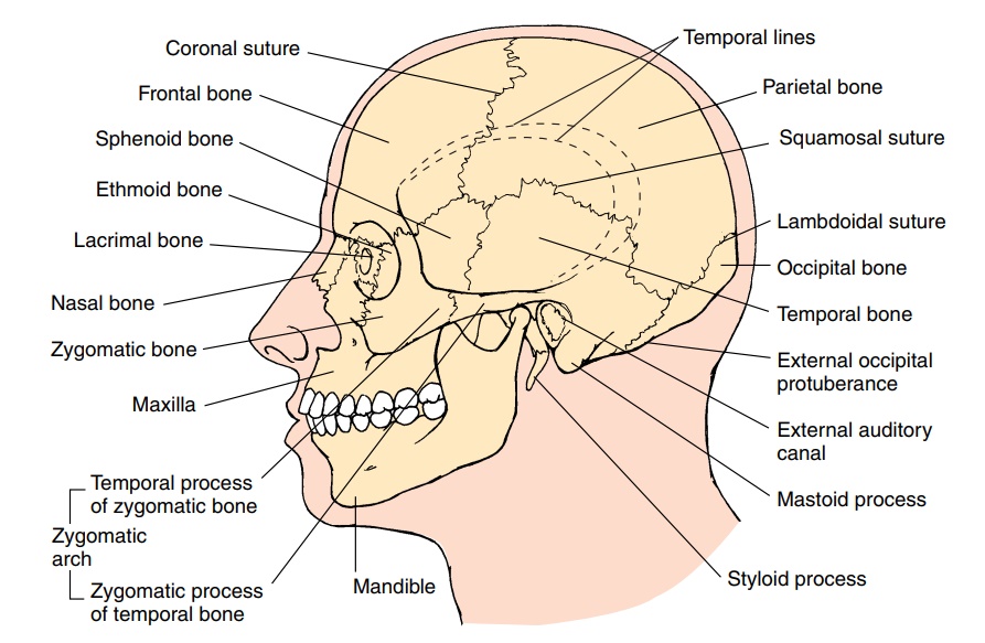
BONES OF THE FACE
The bones of the face include the maxillae, palatines,nasals, mandible, zygomatics, lacrimals, inferior conchae, and vomer.
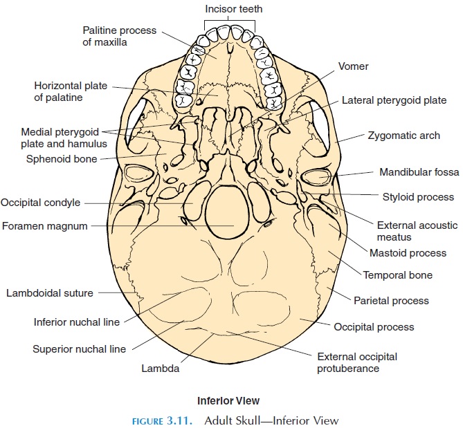
The right and the left maxillae are large facial bones that form the upper jaw. This bone articulates with all the facial bones except the mandible. An im-portant part of this bone is the air-filled cavities, the maxillary sinus, that open into the nasal cavity. Theinferior part of the maxilla forms the alveolar process (Figure 3.9B), which contains the alveoli (sockets) for teeth. A horizontal projection, the palatine process (Figure 3.11), forms the anterior two-thirds of the hard palate (hard part of the roof of mouth).
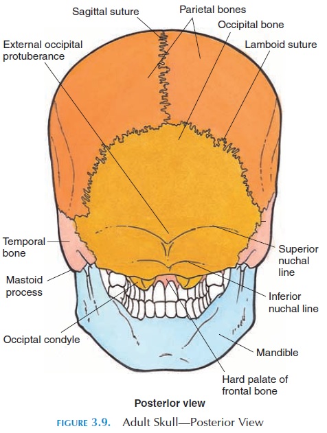
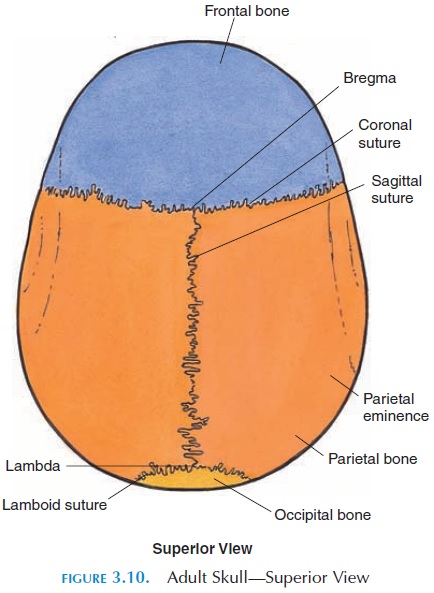
The palatine bones are L-shaped and form the posterior part of the hard palate. The small nasalbones support the superior portion of the bridge ofthe nose. The flexible, cartilaginous portion of the nose is attached to the nasal bone. The vomer is a small bone that forms the inferior portion of the nasal septum (Figure 3.12B). The inferior nasal con-chae (Figure 3.12A) are the lowermost bony projec-tions into the nasal cavity. They have the same func-tion as the middle and superior conchae.
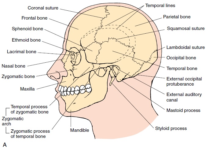
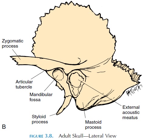
The zygomaticbones, or cheekbones (Figure 3.8), articulates with the frontal, maxilla, sphenoid, and temporal bones. The temporal process articulates with the zygomatic process of the temporal bone, the frontal process with the frontal bone, and the max-illary process with the maxillary bone. This boneforms part of the lateral wall of the orbital cavity. The lacrimal bones (Figure 3.8), the smallest facialbones, are located close to the medial part of the or-bital cavity. They have a lacrimal canal—a small passage that surrounds the tear duct, through which the tears flow from the eye into the nasal cavity. This is why you blow your nose every time you cry.
Related Topics