Milestones in Virology, Size and shape, Characteristic features, Classification, Bacteriophage, Multiplication or Life Cycle of Phages, Viral diseases - Viruses | 11th Botany : Chapter 1 : Living World
Chapter: 11th Botany : Chapter 1 : Living World
Viruses
Viruses
Did you go through the headlines of newspapers in
recent times? Have you heard of the terms EBOLA, ZIKA, AIDS, SARS, H1N1 etc.?
There are serious entities which are considered as “Biological Puzzle” and cause disease in man. They are called
viruses. We have learnt about the attributes of living world in the previous
chapter. Now we shall discuss about viruses which connect the living and
nonliving world.
The word virus is derived from Latin meaning
‘Poison’. Viruses are sub-microscopic, obligate intracellular parasites. They
have nucleic acid core surrounded by protein coat. Viruses in their native
state contain only a single type of nucleic acid which may be either DNA or
RNA. The study of viruses is called Virology.

1. Milestones in Virology
1796 Edward Jenner used vaccination for small pox
1886 Adolf Mayer demonstrated the infectious nature
of Tobacco mosaic virus using sap of mosaic leaves
1898 M.W. Beijierink defined the infectious agent
in tobacco leaves as ῾Contagium vivum
fluidum’
1915 F.W.Twort identified Viral infection in
Bacteria
1917 d’Herelle coined the term ‘Bacteriophage’
1984 Luc Montagnier and Robert Gallo discovered HIV
(Human Immuno Deficiency Virus).
2. Size and shape
Viruses are ultramicroscopic particles. They are
smaller than bacteria and their diameter range from 20 to 300 nm. (1nm =
10-9metres). Bacteriophage measures about 10-100 nm in size. The size of TMV is
300×20 nm.
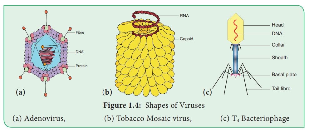
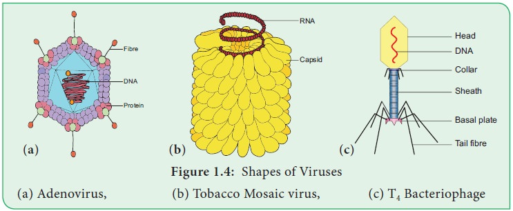
Generally viruses are of three types based on shape
and symmetry (Figure 1.4).
i. Cuboid symmetry – Example: Adenovirus,
Herpes virus.
ii. Helical symmetry – Example: Influenza virus,
TMV.
iii. Complex or Atypical – Example: Bacteriophage,
Vaccinia virus.
3. Characteristic features of Viruses
Living Characters
•
Presence of nucleic acid and protein.
•
Capable of mutation
•
Ability to multiply within living cells.
•
Able to infect and cause diseases in living beings.
•
Show irritability.
•
Host –specific
Non-living Characters
•
Can be crystallized.
•
Absence of metabolism.
•
Inactive outside the host.
•
Do not show functional autonomy.
•
Energy producing enzyme system is absent.
4. Classification of Viruses
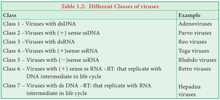
Among various classifications proposed for viruses the classification given by David Baltimore in the year 1971 is given below. The classification is based on mechanism of RNA production, the nature of the genome (single stranded –ss or double stranded - ds ), RNA or DNA, the use of reverse transcriptase(RT), ss RNA may be (1) sense or (2) antisense. Viruses are classified into seven classes (Table 1.2).
Viral genome
Each virus possesses only one type of nucleic acid
either DNA or RNA. The nucleic acid may be in a linear or circular form.
Generally nucleic acid is present as a single unit but in wound tumour virus
and in influenza virus it is found in segments. The viruses possessing DNA are
called ‘Deoxyviruses’ whereas those
possessing RNA are called ‘Riboviruses’ .
Majority of animal and bacterial
viruses are DNA viruses (HIV is the animal virus which possess RNA). Plant
viruses generally contain RNA (Cauliflower Mosaic virus possess DNA). The
nucleic acids may be single stranded or double stranded. On the basis of nature
of nucleic acid viruses are classified into four Categories. They are Viruses
with ssDNA (Parvoviruses), dsDNA (Bacteriophages), ssRNA (TMV) and dsRNA(wound
tumour virus).
5. Tobacco Mosaic Virus (TMV)
Tobacco mosaic virus was discovered in 1892 by
Dimitry Ivanowsky from the Tobacco plant. Viruses infect healthy plants through
vectors like aphids, locusts etc. The first visible symptom of TMV is
discoloration of leaf colour along the veins and show typical yellow and green
mottling which is the mosaic symptom. The downward curling and distortion of
young apical leaves occurs, plant becomes stunted and yield is affected.
Structure
Electron microscopic studies have revealed that TMV
is a rod shaped (Figure 1.4b) helical virus measuring about 280x150µm with a
molecular weight of 39x106 Daltons. The virion is made up of two constituents,
a protein coat called capsid and a
core called nucleic acid. The
protein coat is made up of
approximately 2130 identical protein subunits called capsomeres which are present around a central single stranded RNA
molecule. The genetic information necessary for the formation of a complete TMV
particle is contained in its RNA. The RNA consists of 6,500 nucleotides.
6. Bacteriophage
Viruses
infecting bacteria are called Bacteriophages
. It literally means ‘eaters of
bacteria’ (Gr: Phagein = to eat). Phages are abundant in soil, sewage water,
fruits, vegetables, and milk.
Structure of T4 bacteriophage
The T4 phage is tadpole shaped and
consists of head, collar, tail, base plate and fibres (Figure 1.4). The head is
hexagonal which consists of about 2000 identical protein subunits. The long
helical tail consists of an inner tubular core which is connected to the head
by a collar. There is a base plate attached to the end of tail. The base plate
contains six spikes and tail fibres. These fibres are used to attach the phage
on the cell wall of bacterial host during replication. A dsDNA molecule of
about 50 µm is tightly packed inside the head. The DNA is about 1000 times
longer than the phage itself.
7. Multiplication or Life Cycle of Phages
Phages multiply through two different types of life
cycle. a. Lytic or Virulent cycle b. Lysogenic or Avirulent life cycle
a. Lytic Cycle
During lytic cycle of phage, disintegration of host
bacterial cell occurs and the progeny virions are released (Figure 1.5a). The
steps involved in the lytic cycle are as follows:
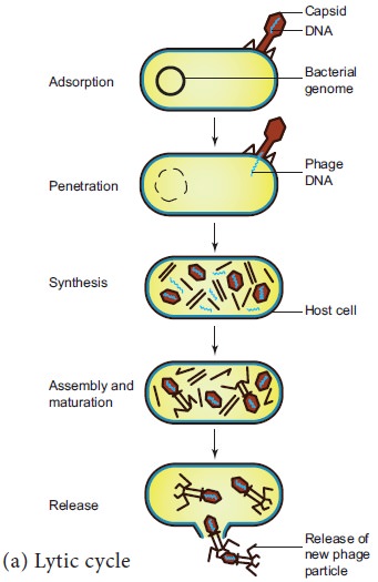
(i)
Adsorption
Phage (T4) particles interact with cell
wall of host (E. coli). The phage
tail makes contact between the two, and tail fibres recognize the specific
receptor sites present on bacterial cell surface. The lipopolysaccharides of
tail fibres act as receptor in phages. The process involving the recognition of
phage to bacterium is called landing.
Once the contact is established between tail fibres and bacterial cell, tail
fibres bend to anchor the pins and base plate to the cell surface. This step is
called pinning.
(ii)
Penetration
The penetration process involves mechani-cal and
enzymatic digestion of the cell wall of the host. At the recognition site phage
digests certain cell wall structure by viral enzyme (lysozyme). After pinning
the tail sheath contracts (using ATP) and appears shorter and thicker. After
contraction of the base plate enlarges through which DNA is injected into the
cell wall without using metabolic energy. The step involving injection of DNA
particle alone into the bacterial cell is called Transfection. The empty protein coat leaving outside the cell is
known as ‘ghost’.
(iii)
Synthesis
This step involves the degradation of bacterial
chromosome, protein synthesis and DNA replication. The phage nucleic acid takes
over the host biosynthetic machinery. Host DNA gets inactivated and breaks
down. Phage DNA suppresses the synthesis of bacterial protein and directs the
metabolism of the cell to synthesis the proteins of the phage particles and
simultaneously replication of Phage DNA also takes place.
The DNA of the phage and protein coat are
synthesized separately and are assembled to form phage particles. The process
of
Figure
1.5: Multiplication cycle of phage,
![]()
![]()
![]()
(v)
Release
The phage particle gets accumulated inside the host
cell and are released by the lysis of host cell wall.
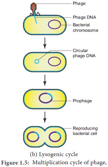
b. Lysogenic Cycle
In the lysogenic cycle the phage DNA gets
integrated into host DNA and gets multiplied along with nucleic acid of the
host. No independent viral particle is formed (Figure 1.5b).
As soon as the phage injects its linear DNA into
the host cell, it becomes circular and integrates into the bacterial chromosome
by recombination. The integrated phage DNA is now called prophage. The activity of the prophage gene is repressed by two repressor proteins which are synthesized
by phage genes. This checks the synthesis of new phages within the host cell.
However, each time the bacterial cell divides, the prophage multiplies along
with the bacterial chromosome. On exposure to UV radiation and chemicals the
excision of phage DNA may occur and results in lytic cycle.
Virion is an
intact infective virus particle
which is non-replicating outside a host cell.
Viroid is a
circular molecule of ssRNA without a
capsid and was discovered by T.O.Diener in the year 1971. The RNA of viroid has
low molecular weight. Viroids cause citrus exocortis and potato spindle tuber
disease in plants.
They are the small circular RNAs which are similar
to viroids but they are always linked with larger molecules of the viral RNA.
Prions were
discovered by Stanley B. Prusiner in
the year 1982 and are pro-teinaceous infectious particles. They are the
causative agents for about a dozen fatal degenerative disorders of the
central nervous system of humans and other animals . For example Creutzfeldt – Jakob
Disease (CJD), Bovine Spongiform En-cephalopathy (BSE) – commonly known as mad
cow disease and scrapie disease of sheep.
Viruses infecting blue green algae are called Cyanophages and are first reported by
Safferman and Morris in the year 1963(Example LPP1 - Lyngbya, Plectonema and Phormidium). Similarly, Hollings(1962)
reported viruses infecting cultivated Mushrooms and causing die back disease.
The viruses attacking fungi are called Mycoviruses
or Mycophages.
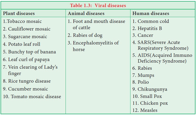
8. Viral diseases
Viruses are known to cause disease in plants, animals
and Human beings (Figure 1.6). A list of viral disease is given in Table 1.3
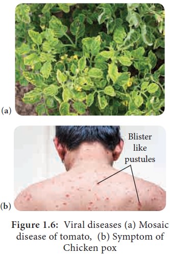
Figure
1.6: Viral diseases (a) Mosaic disease of tomato, (b) Symptom of
Chicken pox
Related Topics