Milestones in Mycology, Characteristic features, Methods of Reproduction, Classification, Economic importance - Fungi | 11th Botany : Chapter 1 : Living World
Chapter: 11th Botany : Chapter 1 : Living World
Fungi
Fungi
![]()
![]()
![]()
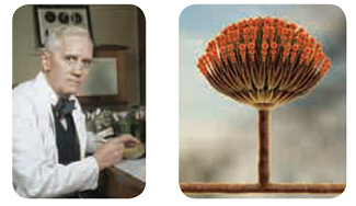
World War II and Penicillin History speaks on fungi
Alexander Fleming
Discovery of Penicillin in the year 1928 is a
serendipity in the world of medicine. The History of World War II recorded the
use of Penicillin in the form of yellow powder to save lives of soldiers. For
this discovery - The wonderful antibiotic he was awarded Nobel Prize in
Medicine in the year 1945.
Milestones in Mycology
1729 P.A.Micheli conducted spore culture
experiments
1767 Fontana proved that Fungi could cause disease
in plants
1873 C.H. Blackley proved fungi could cause allergy
in Human beings
1906 A.F.Blakeslee reported heterothallism in fungi
1952 Pontecarvo and Raper reported Parasexual cycle
The word ‘fungus’ is derived from Latin meaning
‘mushroom’. Fungi are ubiquitous, eukaryotic, achlorophyllous heterotrophic
organisms. They exist in unicellular or multicellular forms. The study of fungi
is called mycology. (Gr. mykes – mushroom: logos – study). P.A. Micheli is
considered as founder of Mycology. Few renowned mycologists include Arthur H.R.
Buller, John Webster,
D.L.Hawksworth, G.C.Ainsworth, B.B.Mundkur, K.C.Mehta, C.V. Subramanian
and T.S. Sadasivan.
E.J. Butler (1874-1943) : Father of Indian
Mycology. He established Imperial Agricultural Research Institute at Pusa,
Bihar. It was later shifted to New Delhi and at present known as Indian
Agricultural Research Insitute (IARI) He published a book, ‘Fungi and Disease
in Plants’ on Indian plant diseases in the year 1918.
General characteristic features
•
Majority of fungi are made up of thin, filamentous
branched structures called hyphae. A number of hyphae get interwoven to form
mycelium. The cell wall of fungi is made up of a polysaccharide called chitin
(polymer of N-acetyl glucosamine).
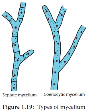
• The fungal mycelium is categorised into two types based on the presence or absence of septa (Figure 1.19). In lower fungi the hypha is aseptate, multinucleate and is known as coenocytic mycelium (Example: Albugo). In higher fungi a septum is present between the cells of the hyphae. Example: Fusarium.
•
The mycelium is organised into loosely or compactly
interwoven fungal tissues called plectenchyma.
It is further divided into two types prosenchyma
and pseudoparenchyma. In the former
type the hyphae are arranged loosely but parallel to one another In the latter
hyphae are compactly arranged and loose their identity.
•
In holocarpic forms the entire thallus is converted
into reproductive structure whereas in Eucarpic some regions of the thallus are
involved in the reproduction other regions remain vegetative. Fungi reproduce
both by asexual and sexual methods. The asexual phase is called Anamorph and the sexual phase is called
Teleomorph. Fungi having both phases
are called Holomorph.
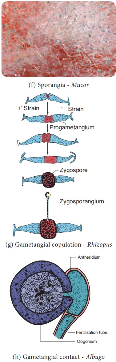
In general sexual reproduction in fungi includes
three steps 1. Fusion of two protoplasts (plasmogamy) 2. Fusion of nuclei
(karyogamy) and 3. Production of haploid spores through meiosis. Methods of
reproduction in fungi is given in Figure 1.20.
Methods of Reproduction in Fungi
Asexual Reproduction
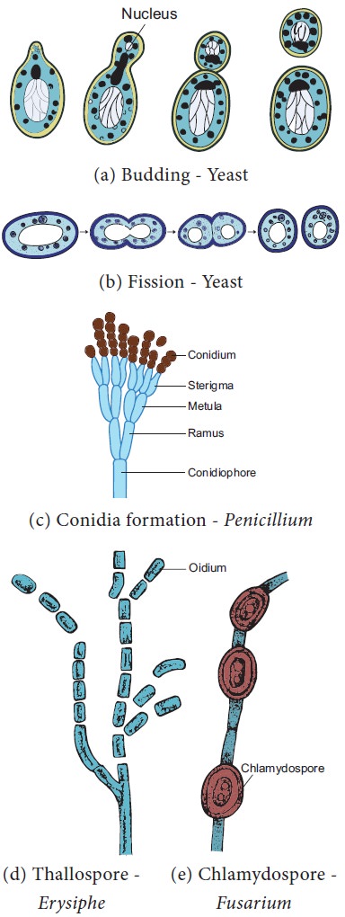
1. Zoospores:
They are flagellate structures produced in zoosporangia (Example: Chytrids)
2. Conidia:
The spores produced on condiophores (Example:
Aspergillus)
3. Oidia/Thallospores/Arthrospores:
The hypha divide and develop in to spores called oidia (Example: Erysiphe).
4. Fission:
The vegetative cell divide into 2 daughter cells. (Example: Schizosaccharomyces-yeast).
5.
Budding: A small outgrowth is developed on parent
cell, which gets detached and become independent. (Example: Saccharomyces-yeast)
6. Chlamydospore:
Thick walled resting spores are called chlamydospores (Example: Fusarium).
Sexual Reproduction
1.Planogametic
copulation: Fusion of motile gamete is called planogametic copulation. a.
Isogamy – Fusion of morphologically and physiologicall similar gametes.
(Example: Synchytrium). b. Anisogamy
– Fusion of morphologically or
physiologically dissimilar gametes (Example: Allomyces). c. Oogamy – Fusion of both morphologi-cally and
physiologically dissimilar gam-etes. (Example: Monoblepharis)
2.
Gametangial contact: During sexual reproduction a
contact is established between antheridium and Oogonium (Example: Albugo)
3. Gametangial copulation: Fusion of gametangia to
form zygospore (Example: Mucor, Rhizopus).
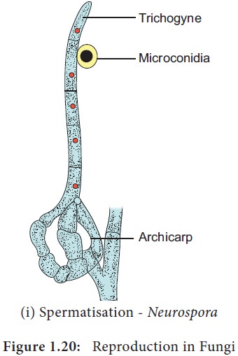
4. Spermatization:
In this method a un-inucleate pycniospore/microconidium is transferred to
receptive hyphal cell (Example: Puccinia/Neurospora)
5. Somatogamy: Fusion of two somatic cells of the hyphae (Example: Agaricus)
Classification of Fungi
Many mycologists have attempted to classify fungi
based on vegetative and reproductive characters. Traditional classifications
categorise fungi into 4 classes – Phycomycetes, Ascomycetes, Basidiomycetes and
Deuteromycetes. Among these ‘Phycomycetes’ include fungal species of Oomycetes,
Chytridiomycetes and Zygomycetes which are considered as lower fungi indicating
algal origin of fungi. Constantine J. Alexopoulos and Charles W. Mims in the
year 1979 proposed the classification of fungi in the book entitled
‘Introductory Mycology’. They classified fungi into three divisions namely
Gymnomycota, Mastigomycota and Amastigomycota. There are 8 subdivisions, 11
classes, 1 form class and 3 form subclasses in the classification proposed by
them.
The
outline of the classification is given below:
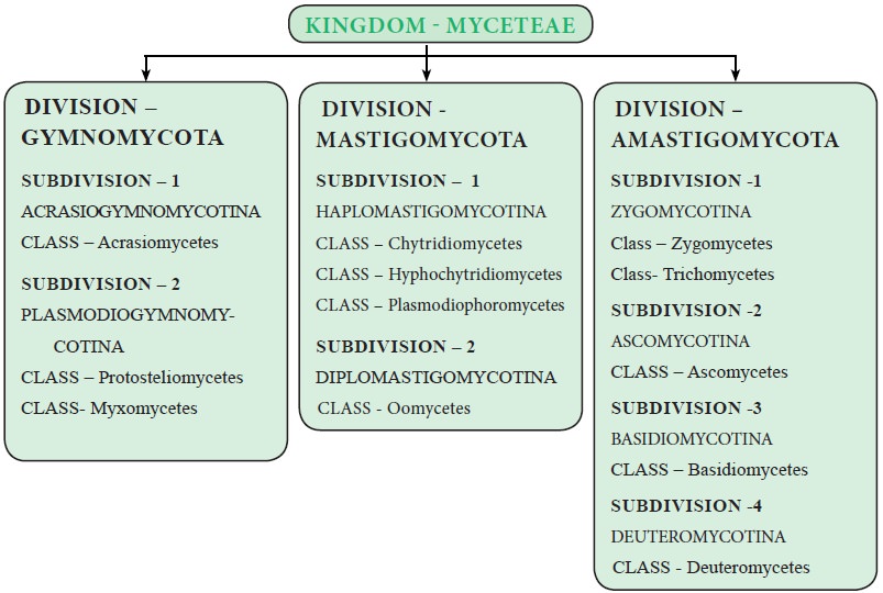
Kingdom : Myceteae (Fungi)
Include achlorophyllous, saprophytic or parasitic
organisms with Unicellular or multicellular (Mycelium) thallus surrounded by
chitinous cell wall. Nutrition is absorptive except slime molds.Reproduction is
through asexual and Sexual methods.
Division : I Gymnomycota
Nutrition Phagotrophic, members of this group lack
cell wall. Example. Dictyostelium
Division :II Mastigomycota
Flagellate
cells are present(Gamete/ Zoospore) . Nutrition
absorptive, mycelium coenocytic. Example : Albugo
Division : III Amastigomycota
Unicellular to multicellular forms are included.
The mycelium is septate.
Asexual reproduction occurs by budding,
fragmentation, sporangiospores, conidia etc., Meiosis is zygotic. Example : Peziza
Recently, with the advent of molecular methods
myxomycetes and oomycetes were reclassified and treated under chromista.
The salient features of some of the classes –
Oomycetes, Zygomycetes, Ascomycetes, Basidiomycetes and Form class
Deuteromycetes are discussed below.
Oomycetes
Coenocytic mycelium is present. The cell wall is
made up of Glucan and Cellulose. Zoospore with one whiplash and one tinsel
flagellum is present. Sexual reproduction is Oogamous. Example: Albugo.
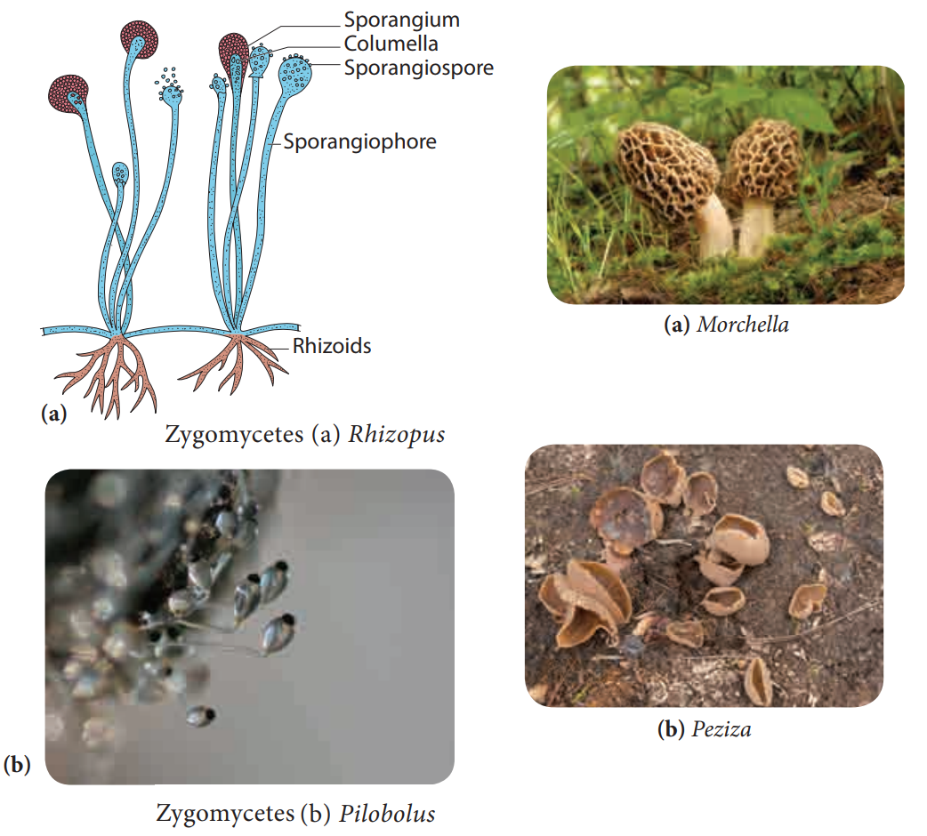
Zygomycetes
•
Most of the species are saprophytic and live on
decaying plant and animal matter in the soil. Some lead parasitic life
(Example: Entomophthora on housefly)
•
Bread mold fungi (Example: Mucor, Rhizopus) and
Coprophilous fungi (Fungi growing on
dung Example: Pilobolus) belong to
this group (Figure 1.21).
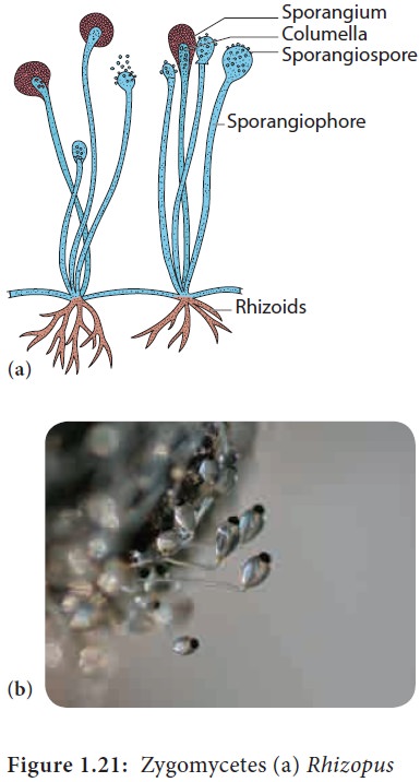
• The mycelium is branched and coenocytic
•
Asexual reproduction by means of spores produced in
sporangia.
•
Sexual reproduction is by the fusion of the
gametangia which results in thick walled zygospore. It remains dormant for long
periods. The zygospore undergoes meiosis and produce spores.
![]()
![]()
![]()
Ascomycetes
• Ascomycetes
include a wide
range of fungi such as yeasts, powdery mildews, cup fungi, morels and so
on (Figure 1.22).
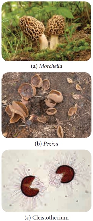
•
Although majority of the species live in
terrestrial environment, some live in aquatic environments both fresh water and
marine.
•
The mycelium is well developed, branched with
simple septum.
•
Majority of them are saprophytes but few parasites
are also known (Powdery mildew – Erysiphe).
•
Asexual reproduction takes place by fission,
budding, oidia, conidia, chlamydospore.
•
Sexual reproduction takes place by the fusion of
two compatible nuclei.
•
Plasmogamy is not immediately followed by
karyogamy, instead a dikaryotic condition is prolonged for several generations.
•
A special hyphae called ascogenous hyphae is
formed.
•
A crozier is formed when the tip of the ascogenous
hyphae recurves forming a hooked cell. The two nuclei in the penultimate cell
of the hypha fuse to form a diploid nucleus. This cell form young ascus.
•
The diploid nucleus undergo meiotic division to
produce four haploid nuclei, which further divide mitotically to form eight
nuclei. The nucleus gets organised into 8 ascospores.
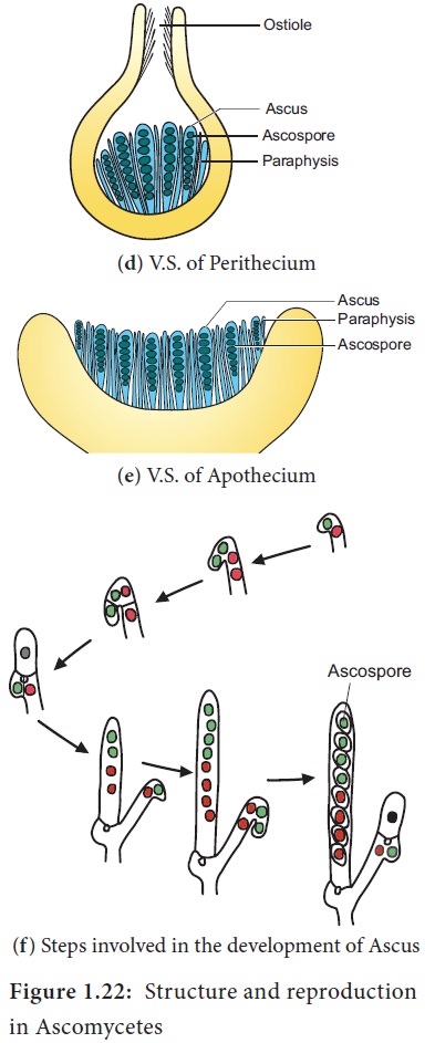
•
The ascospores are found inside a bag like
structure called ascus. Due to the presence of ascus, this group is popularly
called "Sac fungi".
•
Asci gets surrounded by sterile hyphae forming
fruit body called ascocarp.
•
There are 4 types of ascocarps namely Cleistothecium (Completely closed), Perithecium (Flask shaped with ostiole), Apothecium (Cup shaped, open type) and Pseudothecium.
Basidiomycetes
•
Basidiomycetes include puff balls, toad stools,
Bird’s nest fungi, Bracket fungi, stink horns, rusts and smuts (Figure 1.23).
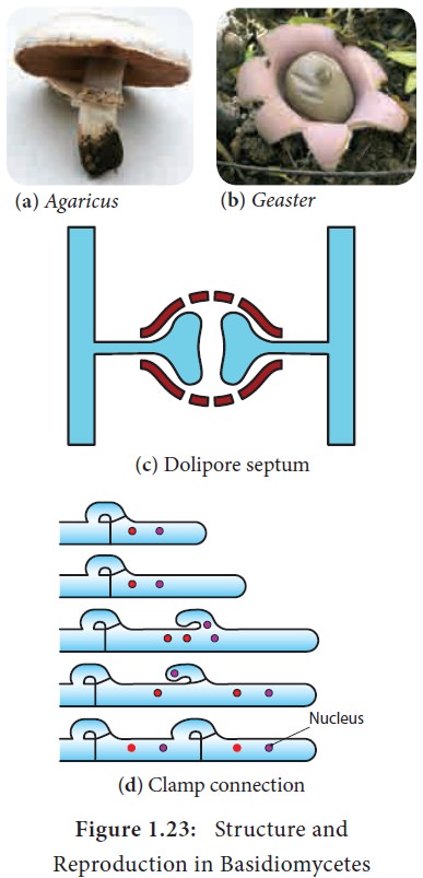
•
The members are terrestrial and lead a saprophytic
and parasitic mode of life.
•
The mycelium is well developed, septate with
dolipore septum(bracket like). Three types of mycelium namely Primary
(Monokaryotic), Secondary (Dikaryotic) and tertiary are found.
• Clamp connections are formed to maintain dikaryotic condition.
•
Asexual reproduction is by means of conidia, oidia
or budding.
•
Sexual reproduction is present but sex organs are
absent. Somatogamy or spermatisation results in plasmogamy. Karyogamy is
delayed and dikaryotic phase is prolonged. Karyogamy takes place in basidium
and it is immediately followed by meiotic division.
•
The four nuclei thus formed are transformed into
basidiospores which are borne on sterigmata outside the basidium (Exogenous ).
The basidium is club shaped with four basidiospores, thus this group of fungi
is popularly called “Club fungi”. The fruit body formed is called Basidiocarp.
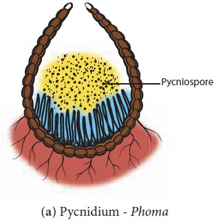
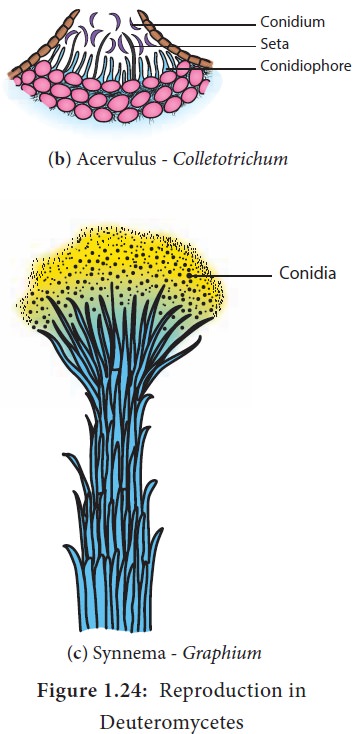
Deuteromycetes or Fungi Imperfecti
The fungi belonging to this group lack sexual reproduction and are called imperfect fungi. A large number of species live as saprophytes in soil and many are plant and animal parasites. Asexual reproduction takes place by the production of conidia, chlamydospores, budding, oidia etc., Conidia are also produced in special structures called pycnidium, Acervulus, sporodochium and Synnema (Figure 1.24). Parasexual cycle operates in this group of fungi. This brings genetic variation among the species.
Economic importance
Fungi provide delicious and nutritious food called
mushrooms. They recycle the minerals by decomposing the litter thus adding
fertility to the soil. Dairy industry is based on a single celled fungus called
yeast. They deteriorate the timber. Fungi cause food poisoning due the
production of toxins. The Beneficial and harmful activities of fungi are
discussed below:
Beneficial activities
![]()
![]()
![]()
Food
Mushrooms like Lentinus
edodes, Agaricus bisporus,
Volvariella volvaceae are consumed for their high nutritive value. Yeasts
provide vitamin B and Eremothe-cium
ashbyii is a rich source of Vitamin B12.
Medicine
Fungi produce antibiotics which arrest the growth
or destroy the bacteria. Some of the antibiotics produced by fungi include
Penicillin (Penicillium notatum)
Cephalosporins (Acremonium chrysogenum) Griseofulvin (Penicillium griseofulvum). Ergot
alkaloids (Ergota-mine) produced by Claviceps
purpurea is used as vasoconstrictors.
Industries
Production
of Organic acid: For the commercial
production of organic acids fungi are employed in the Industries. Some of the
organic acids and fungi which help in the production of organic acids are:
Citric acid and Gluconic acid – Aspergillus
niger, Itaconic acid – Aspergillus terreus, Kojic acid – Aspergillus oryzae
Bakery and Brewery
Yeast(Saccharomyces
cerevisiae) is used for fermentation of sugars to yield alcohol. Bakeries
utilize yeast for the production of Bakery products like Bread, buns, rolls
etc., Penicillium roquefortii and Penicillium camemberti were employed in cheese production.
Production of enzymes
Aspergillus
oryzae, Aspergillus niger were
employed in the production of enzymes like Amylase, Protease, Lactase etc.,’
Rennet’ which helps in the coagulation of milk in cheese manufacturing is
derived from Mucor spp.
Agriculture
Mycorrhiza forming fungi like Rhizoctonia, Phallus,
Scleroderma helps in absorption
of water and minerals.
Fungi like Beauveria
bassiana, Metarhizium anisopliae are
used as Biopesticides to eradicate
the pests of crops.
Gibberellin, produced by a fungus Gibberella fujikuroi induce the plant growth and is used as growth promoter.
Harmful activities
![]()
![]()
![]()
Fungi like Amanita
phalloides , Amanita verna, Boletus satanus are highly poisonous due to the production of Toxins. These fungi are commonly referred
as “Toad stools”.
Aspergillus
, Rhizopus, Mucor and Penicilium are involved in spoilage of food materials. Aspergillus
flavus infest dried foods and produce carcinogenic toxin called aflatoxin.
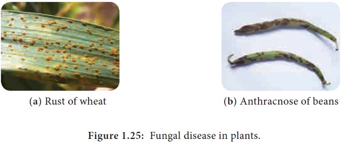
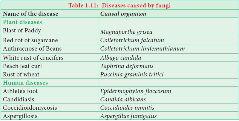
Fungi
cause diseases in Human beings and Plants (Table 1.11 and Figure 1.25)
Activity 1.4
Get
a button mushroom. Draw diagram of the fruit body. Take a thin longitudinal
section passing through the gill and observe the section under a microscope.
Record your observations.
Activity 1.5
Keep
a slice of bread in a clean plastic tray or plate. Wet the surface with little
water. Leave the setup for 3 or 4 days. Observe the mouldy growth on the
surface of the bread. Using a needle remove some mycelium and place it on a
slide and stain the mycelium using lactophenol cotton blue. Observe the
mycelium and sporangium under the microscope and Record your observation and
identify the fungi and its group based on characteristic features.
Rhizopus
Class - Zygomycetes
Order - Mucorales
Family - Mucoraceae
Genus - Rhizopus
Rhizopus is a
saprophytic fungus and grows on
substrates like bread, jelly, leather, decaying vegetables and fruits. It is
commonly called ‘Bread mold’. Rhizopus
stolonifer causes leak and soft rot
of vegetables
![]()
![]()
![]()
Vegetative structure
The mycelium consists of aseptate, multinucleate (coenocyte) and profusely branched hyphae. There are horizontally growing aerial hyphae called stolons. The stolons produce rhizoids which are branched and penetrate the substratum and help in absorbing water and nutrients. Sporangiophores are borne exactly opposite to the rhizoids. The cell wall is made up of chitin and chitosan. The cell wall is followed by plasma membrane. The protoplast is granular containing many nuclei. Cell organelles like mitochondria, ribosomes and endoplasmic reticulum are present. The cell inclusions like glycogen and oil droplets are also found.
Reproduction
Rhizopus reproduces by asexual and
sexual methods.
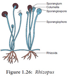
Asexual reproduction
During favorable conditions, erect sporangiophores
are produced exactly opposite to the region of formation of rhizoids of the
mycelium. The sporangiophores are unicellular, unbranched and multinucleate
structures which bear bag like structure called sporangia. Each sporangiophore
bears a single sporangium.
Sporangium possesses a sterile region in the centre
called Columella. Spores are
produced around the columella. When the sporangial wall breaks, the columella
collapses and the spores are dispersed. When the spores fall on a suitable
substratum they germinate and produce new mycelia (Figure 1.26).
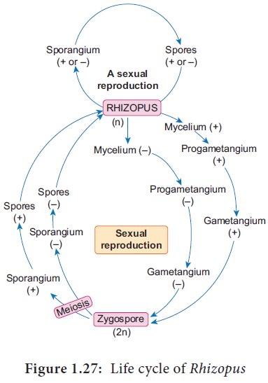
Sexual reproduction
Sexual reproduction is present and takes place
through gametangial copulation. Most of the species are heterothallic but Rhizopus sexualis is homothallic. There
is no morphological distinction between the two sexual hyphae although
physiologically they are dissimilar. Since physiologically dissimilar thalli (hyphae)
are involved in sexual reproduction, this phenomenon is called heterothallism. Mycelia which produce
gametangia are of opposite strains (+) or (-). The first step is the formation
of special hyphae called zygophores. The tips of the two zygophores swell to
form progametangia. Further, a septum is formed near the tip of each
progametangium and results in the formation of a terminal gametangium and a
suspensor cell. The two gametangia fuse, and this is followed by plasmogamy and
karyogamy. The fusion of nuclei results in the formation of a diploid
zygospore. Many nuclei belonging to opposite strains (+ or –) pair and fuse to
form many diploid nuclei. The zygospore enlarges and develops an outer thick
dark and warty layer called exine and inner thin layer called intine. After the
resting period the nuclei of zygospore undergo meiosis. The zygospore
germinates to form sporangiophores and the zygosporangium contain mixture of
(+)and (–) spores. When the spores fall on a suitable substratum, they germinate
to produce mycelium (Figure 1.20). The life cycle of Rhizopus is given in figure 1.27.
Agaricus
Class - Basidiomycetes
Order - Agaricales
Family - Agaricaceae
Genus - Agaricus
It is a saprophytic fungus found on wood logs, manure
piles, fresh litter, pastures etc., The fruit bodies are the visible part of
the fungi. They are found in rings in some species like Agaricus arvensis, Agaricus
tabularis and hence popularly called
‘Fairy rings”. Agaricus campestris is
the most common ‘field mushroom’.
Vegetative structure
The thallus is made up of branched structures
called hyphae. A large number of hyphae constitute the mycelium.
Three types of mycelia are seen namely primary
mycelium, secondary mycelium and tertiary mycelium, The primary mycelium
develops from the germination of basidiospore. It is septate, uninucleate and
haploid. It is also called monokaryotic
mycelium. Fusion of two primary
mycelium of opposite strains give rise to secondary mycelium or dikaryotic mycelium. The dikaryotic mycelium develops into hyphal cords called Rhizomorphs,. and perennates the soil for a long period. The tertiary mycelium is found in the fruit
body called basidiocarp. Each cell
of the hyphae posssess a cell wall made up of chitin and cell organelles like
mitochondria, golgibodies, Endoplasmic reticulum etc., are also present.
Asexual reproduction.
Agaricus produces
chlamydospores during asexual
reproduction. During favourable condition the chlamydospores germinate and produce
mycelium.
Sexual reproduction
Agaricus reproduces
by sexual method but sex organs are
absent.Majority of the species are heterothallic. Agaricus bisporus is a homothallic species. The opposite strains of
mycelium fuse(somatogamy) and results in the formation of dikaryotic or
secondary mycelium. Karyogamy takes place in basidium and it is immediately
followed by meiosis giving rise to four haploid basidiospores. The
basidiospores are borne on sterigmata. The subterranean mycelial strands called
rhizomorphs posssess dense knots of dikaryotic hyphae. These knots develop into
Basidiocarps.
![]()
![]()
![]()
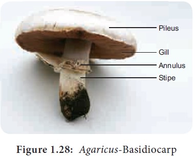
Basidiocarp
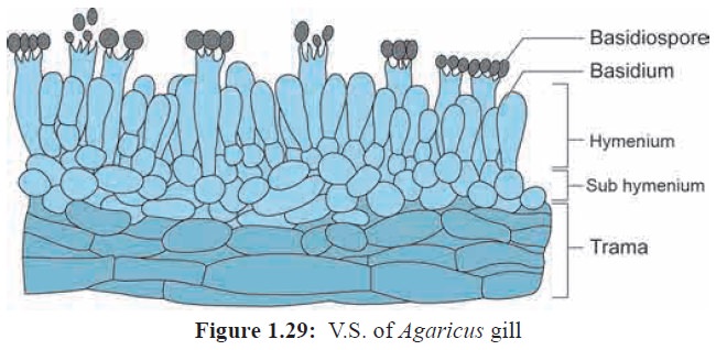
The mature basidiocarp is umbrella shaped and is divided into 3 parts namely stipe, pileus and gill. The stipe is thick, fleshy and cylindrical in structure. The upper part of the stipe possess a membranous structure called annulus. The upper convex surface is called Pileus which is white or cream in colour (Figure 1.28). The inner surface of pileus shows radially arranged gills or lamellae. The gills vary in length. On both the sides of the gills a fertile layer called hymenium is present. The stipe is hollow from the centre and the central part is made up of loosely arranged hyphae whereas the periphery is made up of compactly arranged hyphae forming pseudoparenchymatous tissue. The gill region is divided into 3 regions. The central part of gill between two hymenial layers is called Trama (Figure 1.29). The subhymenial layers have closely compact tissue . The hymenium is the fertile layer and possess club shaped basidia. The basidium is interspersed with sterile hyphae called paraphysis. Each basidium bears 4 basidiospores , of these two basidiospore belong to (+) strain and other two of them will be (–) strain. The basidiospores are borne on stalk like structures called Sterigmata. The basidiospore on germination produces the haploid primary mycelium.
Thus the life cycle of Agaricus shows a very short diploid phase, haploid phase and a
prolonged dikaryotic phase (Figure 1.30).

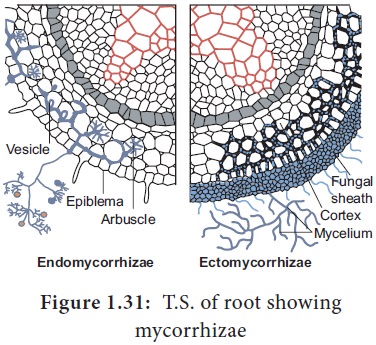
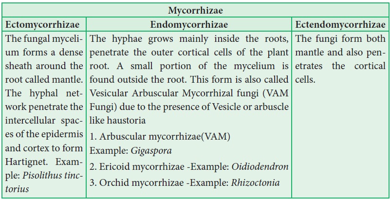
Mycorrhizae
The symbiotic association between fungal mycelium
and roots of plants is called as mycorrhizae. In this relationship fungi
absorbs nutrition from the root and in turn the hyphal network of mycorrhizae
forming fungi helps the plant to absorb water and mineral nutrients from the
soil (Figure 1.31) Mycorrhizae are classified into three types
Importance of Mycorrhizae
•
Helps to derive nutrition in Monotropa, a saprophytic angiosperm,
• Improves the availability of minerals and water to the plants.
•
Provides drought resistance to the plants
•
Protects roots of higher plants from the attack of
plant pathogens
Lichens
The symbiotic association between algae and fungi is called lichens. The algal partner is called Phycobiont or Photobiont., and the fungal partner is called Mycobiont. Algae provide nutrition for fungal partner in turn fungi provide protection and also help to fix the thallus to the substratum through rhizinae.
Asexual reproduction takes place through
fragmentation, Soredia and Isidia. Phycobionts reproduce by akinetes,
hormogonia, aplanospore etc., Mycobionts undergo sexual reproduction and
produce ascocarps.
Classification
•
Based on the habitat lichens are classified into
following types: Corticolous( on
Bark) Lignicolous(on Wood) Saxicolous(on rocks) Terricolous(on ground) Marine(on siliceous rocks of sea) Fresh water(on
siliceous rock of fresh water).
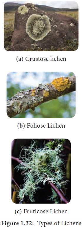
• On the basis of morphology of the thallus they are divided into Leprose (a distinct fungal layer is absent) Crustose-crust like; Foliose-leaf like; Fruticose- branched pendulous shrub like (Figure 1.32).
•
The distribution of algal cells distinguishes
lichens into two forms namely Homoiomerous (Algal cells evenly distributed in
the thallus) and Heteromerous (a distinct layer of algae and fungi present).
• If the fungal partner of lichen belongs to ascomycetes, it is called Ascolichen and if it is basidiomycetes it is called Basidiolichen.
Lichens secrete organic acids like Oxalic acids
which corrodes the rock surface and helps in weathering of rocks, thus acting
as pioneers in Xerosere. Usnic acid produced from lichens show antibiotic
properties. Lichens are sensitive to air pollutants especially to sulphur-
di-oxide. Therefore, they are considered as pollution indicators. The dye
present in litmus paper used as acid base indicator in the laboratories is
obtained from Roccella montagnei.
Cladonia rangiferina (Reindeer
moss) is used as food for animals
living in Tundra regions.
Related Topics