Chapter: 11th Botany : Chapter 1 : Living World
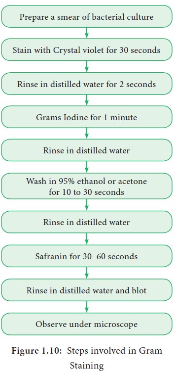
Gram staining procedure - Bacteria
Gram staining procedure
The Gram staining method to differentiate bacteria
was developed by Danish Physician Christian Gram in the year 1884. It is a
differential staining procedure and it classifies bacteria into two classes -
Gram positive and Gram negative. The steps involved in Gram staining procedure
is given in Figure 1.10. The Gram positive bacteria retain crystal violet and
appear dark violet whereas Gram negative type loose the crystal violet and when
counterstained by safranin appear red under a microscope.
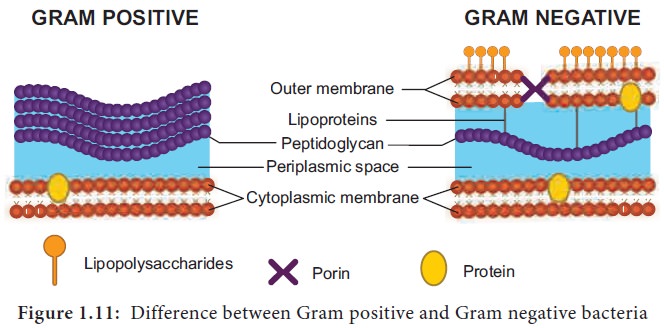
Most of the gram positive cell wall contain
considerable amount of teichoic acid and teichuronic acid. In addition, they
may contain polysaccharide molecules. The gram negative cell wall contains
three components that lie outside the peptidoglycan layer. 1. Lipoprotein 2.
Outer membrane 3.Lipopolysaccharide. Thus the different results in the gram
stain are due to differences in the structure and composition of the cell wall
(Figure 1.11). The difference between Gram Positive and Gram negative bacteria
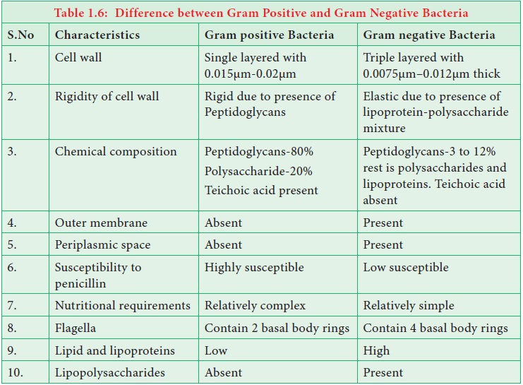
is given in Table 1.6.
What are Magnetosomes ?
Intracellular chains of 40-50 magnetite
(Fe3O4) particles are found in bacterium Aquaspirillum magnetotacticum. and it help the
bacterium to locate nutrient rich sediments.
Related Topics