Botany - Tobacco Mosaic Virus (TMV) and its Structure | 11th Botany : Chapter 1 : Living World
Chapter: 11th Botany : Chapter 1 : Living World
Tobacco Mosaic Virus (TMV) and its Structure
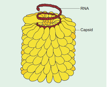
Tobacco Mosaic Virus (TMV)
Tobacco mosaic virus was discovered in 1892 by
Dimitry Ivanowsky from the Tobacco plant. Viruses infect healthy plants through
vectors like aphids, locusts etc. The first visible symptom of TMV is
discoloration of leaf colour along the veins and show typical yellow and green
mottling which is the mosaic symptom. The downward curling and distortion of
young apical leaves occurs, plant becomes stunted and yield is affected.
Structure
Electron microscopic studies have revealed that TMV
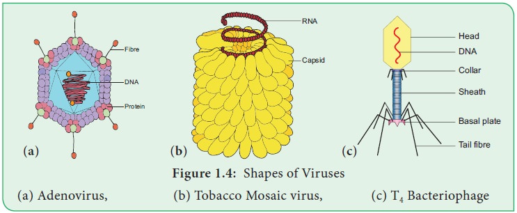
is a rod shaped (Figure 1.4b) helical virus measuring about 280x150µm with a
molecular weight of 39x106 Daltons. The virion is made up of two constituents,
a protein coat called capsid and a
core called nucleic acid. The
protein coat is made up of
approximately 2130 identical protein subunits called capsomeres which are present around a central single stranded RNA
molecule. The genetic information necessary for the formation of a complete TMV
particle is contained in its RNA. The RNA consists of 6,500 nucleotides.
Related Topics