Chapter: Psychology: Sensation
Gathering the Stimulus: The Eye

Gathering the
Stimulus: The Eye
Eyes come in many forms. Some
invertebrates have simple eyespots that merely sense light or dark; others have
complex, multicellular organs with crystalline lenses. In vertebrates, the
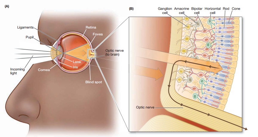
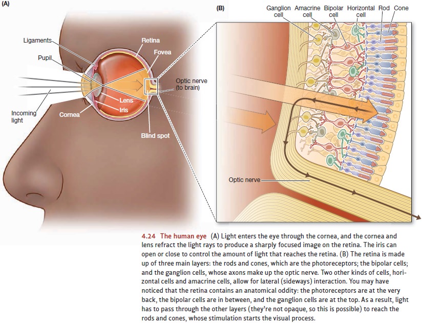
actual detection of light is done by cells called photoreceptors. These cells are located on the retina, a layer of
tissue lining the back of the eyeball. Before the light reaches the retina,
however, several mechanisms are needed to control the amount of light reaching
the photoreceptors and to ensure a clear and sharply focused retinal image.
The iris is a smooth, circular muscle surrounding the pupillary
opening—the open-ing through which light enters the eye. Adjustments in the
iris are under reflex control and cause the pupil to dilate (grow larger) or
contract, thus allowing considerable con-trol over how much light reaches the
retina.
In the mammalian eye, the cornea and the lens focus the incoming light just like a camera lens does (Figure
4.24). The cornea has a fixed shape, but it begins the process of bending the
light rays so they’ll end up properly focused. The fine-tuning is then done by
adjustments of the lens, just behind the cornea. The lens is surrounded by a
ring of ligaments that exert an outward “pull,” causing the lens to flatten
somewhat; this allows the proper focus for objects farther away. To focus on a
nearby object, con-traction of a muscle in the eye reduces the tension on the ligaments
and allows the lens to take on a more spherical shape.

Related Topics