Chapter: Ophthalmology: Cornea
Superficial Punctate Keratitis
Superficial Punctate Keratitis
Definition
Superficial punctate corneal lesions due to
lacrimal system dysfunction from a number causes (see etiology).
Epidemiology and etiology:
Superficial punctate keratoconjunctivitis is avery frequent finding as it can be
caused by a wide variety of exogenous factorssuch as foreign bodies beneath the
upper eyelid, contact lenses, smog, etc. It may also appear as a secondary
symptom of many other forms of keratitis (see the forms of keratitis discussed
in the following section). It can also occur in association with an endogenous
disorder such as Thygeson’s disease.
Symptoms:
Depending on the cause and severity of the
superficial corneallesions, symptoms range from a nearly asymptomatic clinical
course (such as in neuroparalytic keratitis in which the cornea loses its
sensitivity) to an intense foreign body sensation in which the patient has a
sensation of sand in the eye with typical signs of epiphora, severe pain,
burning, and blepharo-spasm. Visual acuity is usually only minimally
compromised.
Diagnostic considerations and differential diagnosis:
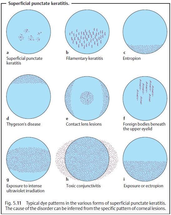
Fluorescein dye isapplied and the eye is
examined under a slit lamp. This visualizes fine epithelial defects. The
specific dye patterns that emerge give the ophthalmol-ogist information about
the etiology of the punctate keratitis (Figs. 5.11a – i).
Treatment and prognosis:
Depending on the cause, the superficial
cornealchanges will respond rapidly or less so to treatment with artificial tears, whereby every effort should be
made to eliminate the causative agents (Fig. 5.11). Depending on the severity of findings, artificial tears of
varying viscos-ity (ranging from eyedrops to high-viscosity gels) are
prescribed and applied with varying frequency. In exposure keratitis, a
high-viscosity gel or ointment is used because of its long retention time;
superficial punctate keratitis is treated with eyedrops.

Related Topics