Chapter: Ophthalmology: Cornea
Corneal Dystrophies
Corneal Dystrophies
Definition
This term refers to a group of corneal
metabolic dysfunctions that always lead to bilateral
opacification of the various layers of the cornea (see Classification below).
Epidemiology:
Corneal dystrophy tends to berare.
The most frequent form isFuchs’ endothelial dystrophy, followed by dystrophy in
the corneal stroma.
Etiology:
The various corneal dystrophies aregenetic disorders. They usuallymanifest themselves in the first or
second decade of life except for Fuchs’ endothelial dystrophy, which only
becomes symptomatic between the ages of 40 and 50.
Classification:
The following forms of dystrophy are differentiated accordingto
the individual layers of the cornea in which they occur:
❖Epithelial corneal dystrophy.
❖ Stromal corneal dystrophy. The most prevalent forms include:
–
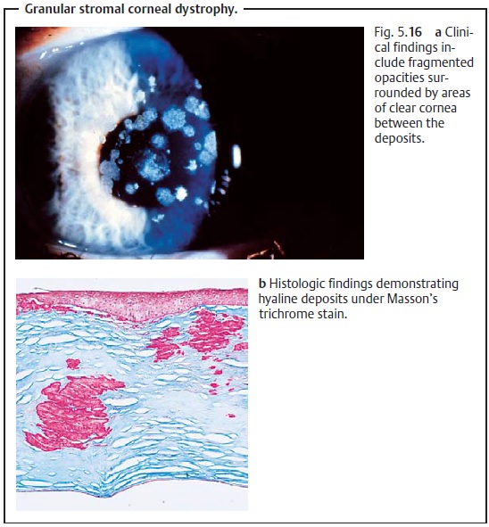
Granular dystrophy (hyaline deposits, Fig. 5.16).
–
Lattice dystrophy (amyloid deposits).
–
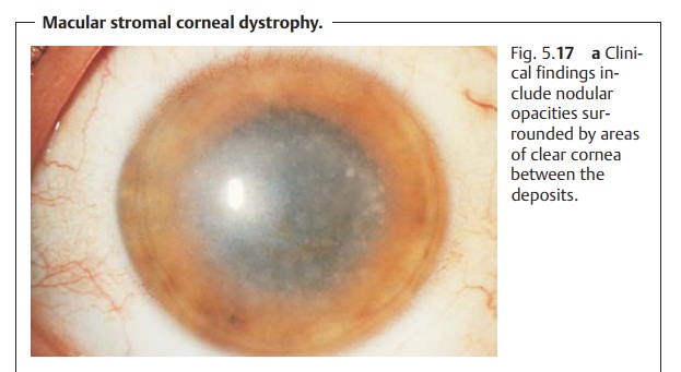
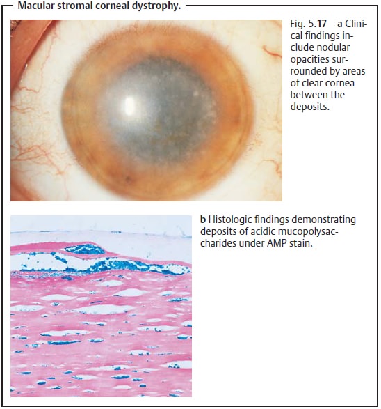
Macular dystrophy (deposits of acidic mucopolysaccharides, Fig. 5.17).
❖ Endothelial dystrophy, such as:
- Fuchs’ endothelial dystrophy (the most
frequently encountered form of corneal dystrophy).
Symptoms and diagnostic considerations:
All patients suffer from asteadily increasing
loss of visual acuity due to the generally gradual opacifica-tion of the
cornea. This loss of visual acuity may progress to the point where a corneal
transplant becomes necessary.
Macular dystrophy is the most rapidly debilitating form of thestromal dys-trophies, resulting in a severe loss of visual acuity in the second decade of life. Epithelial and stromal corneal dystrophies are also often accompanied bypainful and recurrent corneal erosion. Fuchs’ endothelial dystrophy involves a gradual loss of endothelial cells that in time leads to bullous keratopathy (hydration of the cornea with stromal edema and epithelial bullae). The patient typically will have poorer vision in the morning than in the evening, as corneal swelling is greater during the night with the eyelids closed.
Treatment:
Depending on the severity of the loss of visual acuity (see
above),a corneal transplant
(penetrating keratoplasty;) may be indicated. Because the cornea remains
avascular in these disorders, the prognosis is good.
In Fuchs’ endothelial dystrophy, a corneal transplant is the treatment of choice. Where the symptoms are not too far advanced, frequent application ofhyperosmolar solutions can remove water from the cornea. However, this isgenerally only a temporary solution. The corneal transplant is performed in combination with a cataract extraction; patients with Fuchs’ endothelial dystrophy that affects their vision are usually older and also have a cataract. The two procedures are combined because corneal decompensation often results from Fuchs’ endothelial dystrophy following the surgical trauma of cataract extraction.
Related Topics