Chapter: Ophthalmology: Cornea
Bullous Keratopathy

Bullous Keratopathy
Definition
Opacification of the cornea with epithelial
bullae due to loss of function of the endothelial cells.
Epidemiology:
Bullous keratopathy is among the most frequent
indicationsfor corneal transplants.
Etiology:
The transparency of the cornea largely depends
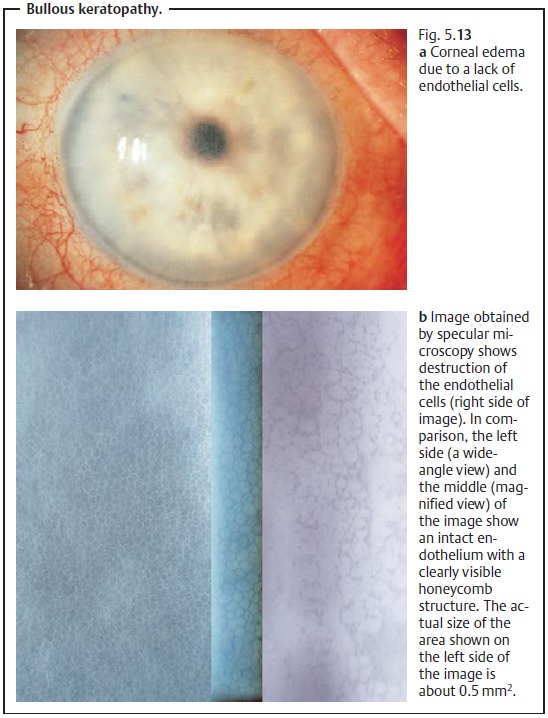
on a functioningendothelium with a high density of endothelial cells (see
Transparency). Where the endothelium has been severely damaged by inflammation,
trauma, or major surgery in the anterior eye, the few remaining endothelial
cells will be unable to prevent aqueous
humor from entering the cornea. This results in hydration of the cornea
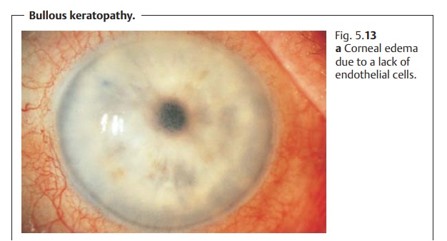
with stromal edema and epithelial bullae (see Figs. 5.13a and b). Loss of
endothelial cells may also have genetic causes (see Fuchs’ endothelial
dystrophy).

Symptoms:
The gradual loss of endothelial cells causesslow deterioration ofvision. The patient
typically will have poorer vision in the morning than in theevening, as corneal
swelling is greater during the night with the eyelids closed.
Diagnostic considerations:
Slit lamp examination will reveal thickening
ofthe cornea, epithelial edema, and epithelial bullae.
Differential diagnosis:
Bullous keratopathy can also occur with
glaucoma.
However, in these cases the intraocular
pressure is typically increased.
Treatment:
Where the damage to the endothelial cells is
not too far advancedand only occasional periods of opacification occur (such as
in the morning), hyperosmolar solutions such
as 5% Adsorbonac can improve the patient’s eye-sight by removing water.
However, this is generally only a temporary solu-tion. Beyond a certain stage a
corneal transplant (penetrating
keratoplasty;) is indicated.
Related Topics