Chapter: Ophthalmology: Cornea
Corneal Deposits

Corneal Deposits
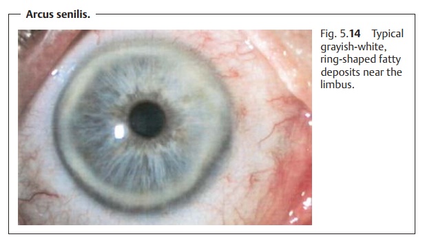
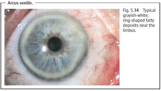
Arcus Senilis
This is a grayish-white ring-shaped
fatty deposit near the limbus that can occur at any age but usually appears in advanced age
(Fig. 5.14). Arcus senilis is usually
bilateral and is a frequently encountered phenomenon. It occurs as a result
of lipid deposits from the vessels of the limbus along the entire periph-ery of
the cornea, which normally increase with advanced age. A lipid-free clear zone
along the limbus will be discernible. Patients younger than 50 years who
develop arcus senilis should be examined to exclude hyper-cholesteremia as a
cause. Arcus senilis requires no
treatment as it does not cause any visual impairments.

The deposits and
pigmentations discussed in the following
section do not generally impair vision.
Corneal Verticillata
Bilateral gray or brownish epithelial deposits that extend in a swirling pattern from a point inferior to the pupil. This corneal change typically occurs with the use of certain medications, most frequently with chloroquine and amiodarone. Fabry’s disease (glycolipid lipidosis) can also exhibit these kinds of corneal changes, which can help to confirm the diagnosis.
Argyrosis and Chrysiasis
Topical medications containing silver and
habitual exposure to silver in elec-troplating occupations lead to silver deposits in the conjunctiva and the
deeplayers of the cornea (argyrosis). Systemic gold therapy (more than 1 – 2 g) willlead to gold coloration of the peripheral corneal
stroma (chrysiasis).
Iron Lines
Any irregularity in the surface of the cornea
causes the eyelid to distribute the tear film irregularly over the surface of
the cornea; a small puddle of tear fluid will be present at the site of the
irregularity. Iron deposits form in a
charac-teristic manner at this site in the corneal epithelium. The most
frequently observed iron lines are the physiologic iron deposits at the site where the eyelids close (the
Hudson-Stähli line), Stocker-Busacca line with pterygium, Ferry’s line with a
filtering bleb after glaucoma surgery, and Fleischer ring with keratoconus.
Iron lines have also been described following surgery (radial keratotomy; photorefractive keratectomy; keratoplasty)
and in thepresence of corneal scars.
Kayser-Fleischer Ring
This golden brown to yellowish
green corneal ring is caused by copper deposits at the level of Descemet’s
membrane in Wilson’s disease (liver and lens degeneration with decreased serum
levels of ceruloplasmin). This ring is so characteristic that the
ophthalmologist often is the first to diagnose this rare clinical syndrome.
Related Topics