Chapter: Psychology: The Brain and the Nervous System
The Synaptic Mechanism
The Synaptic
Mechanism
So far, we’ve suggested that
communication across the synapse (communication between neurons) functions differently from—and is slower than—the
transmission ofinformation within a neuron. But how exactly do neurons
communicate across the synapse? The answer turns out to be chemical—the neuron on the “sending” side of the synapse releases certain
molecules that drift across the synapse and trigger a response on the
“receiving” side. This process is entirely different from the electrical
signaling that takes place within a single neuron.
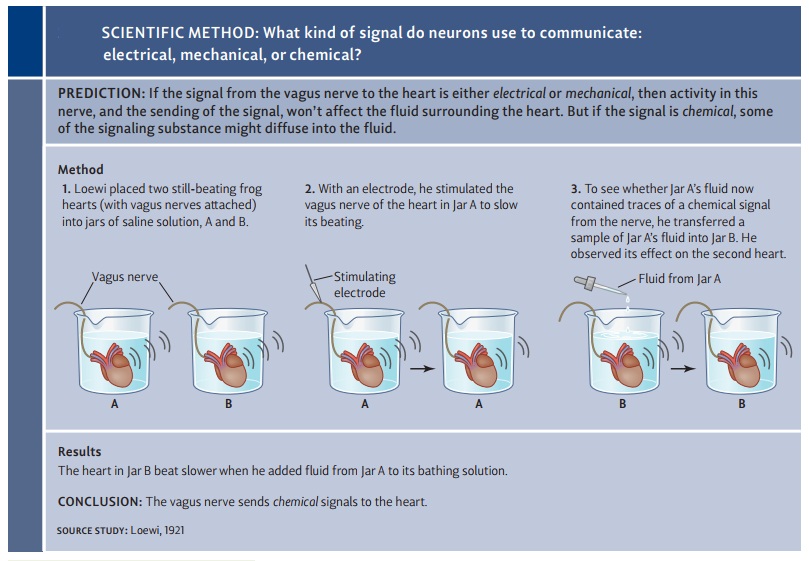
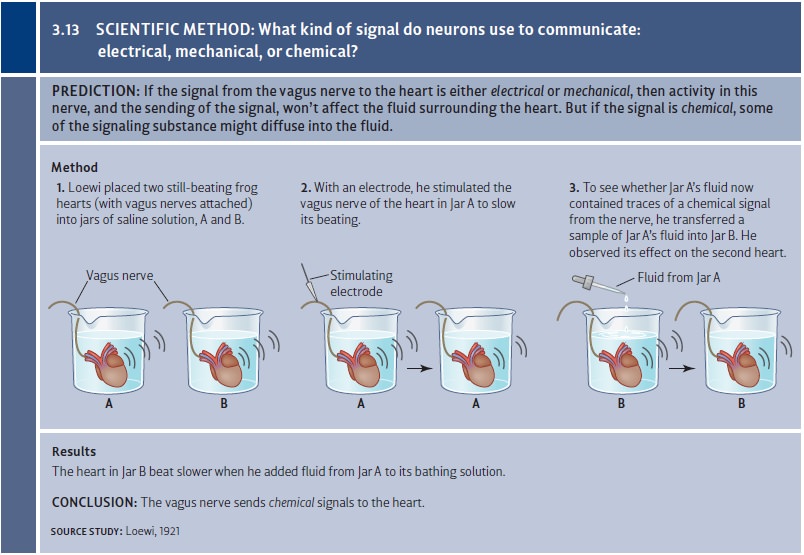
Early insight into these points
came in 1921 from an experiment by Otto Loewi, who later earned the Nobel Prize
for his work. Loewi knew that activity in the vagusnerve causes the heart rate to slow down, and he hypothesized
that the nerve commu-nicates with the heart by releasing a certain chemical. To
test this hypothesis, he dis-sected two frogs and removed their hearts. He
placed each of the hearts, with nerves still attached, in a separate container
filled with fluid. He then electrically stimulated one of the vagus nerves
and—not surprisingly—the attached heart immediately slowed its rate of beating.
Loewi then took a sample of the fluid from that container and dripped it into
the separate container holding the second heart. What should happen? If the
signal from the vagus nerve was electrical or mechanical, then this signal
would not change the fluid in the first container; and so shifting the fluid
into the second container should have
no effect. But if the signal was chemical, then some of the relevant molecules
would probably diffuse out into the fluid of the first con-tainer and be
carried along when the fluid was dripped into the other container. As a result,
the fluid (with these molecules) should slow the second heart, just as it
slowed the first. This is exactly what happened; so apparently, the signal was
chemi-cal (Figure 3.13).
In Loewi’s procedure, the output
from the vagus nerve was a chemical that influenced the muscle tissue in the
heart. In many other contexts, the output from a neuron is a chemical that
triggers a response in another neuron. But how exactly do these chemical
messages work? Let’s focus on what happens when one neuron is sending a signal
to another neuron. The cell that sends the message is called the presynaptic neuron (it’s the one “before
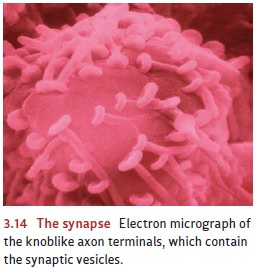
the synapse”), and the actual transmission process begins in the tiny axon terminals of this cell. Within
these swellings are many tiny sacs, or synaptic
vesicles (“little vessels”), that are like water balloons filled with neurotransmitters—the chemicals that
will, when released, influence other neurons.
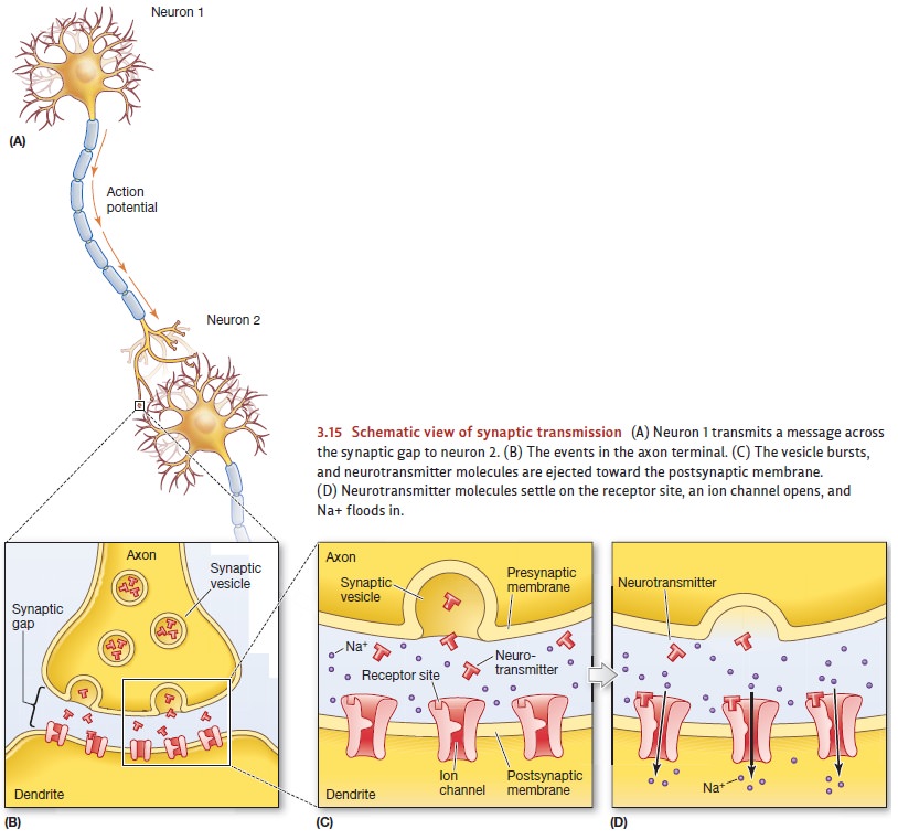
When the presynaptic neuron
fires, some of the vesicles literally burst. They eject their contents into the
gap separating the presynaptic cell from the cell that will receive the
signal—the postsynaptic neuron (the
one “after the synapse”). The neurotransmitter molecules diffuse across this
gap and latch onto receptors on the
membrane of the post-synaptic cell (Figure 3.14; Figure 3.15A and B). This
sequence causes certain ion channels in the postsynaptic membrane to open or
close.
As we’ll see, there are several
types of neurotransmitters, and each type has different effects on the
postsynaptic cell. For example, some neurotransmitters open the chan-nels to
(positively charged) sodium ions (Figure 3.15C and D). This flow of ions will
decrease membrane’s voltage difference, but if the voltage change is small, the
mem-brane may be able to compensate and thus restore the resting potential.
It’s important to realize,
though, that this postsynaptic cell is probably receiving inputs from other
presynaptic cells: They’re also releasing neurotransmitters that latch onto the
cell’s receptors. This activity causes a further flow of sodium into the
postsynaptic neuron, driving its voltage farther and farther from the resting
level. Eventually, the voltage differ-ence for the postsynaptic cell may reach
the cell’s excitation threshold. This triggers an action potential in this cell
that will speed down this neuron’s axon, causing it to release
neurotransmitters of its own.
In other cases, the
neurotransmitters latching onto the receptors can have the opposite effect—they
can make the postsynaptic cell less
likely to fire. Specifically, at some synapses, the presynaptic cell releases
transmitter substances that lead to an some synapses, the presynaptic cell
releases transmitter substances that lead to an increased voltage difference
across the membrane
of the postsynaptic
neuron. This can happen, for example, if the transmitters cause the
opening of channels
that let chloride
ions (Cl") enter the postsynaptic cell. These ions make the inside
of the postsynaptic
cell even more
negatively charged than it
was at the
start. The heightened voltage difference moves the cell away
from its excitation thresh- old, making it less likely to fire.
Most neurons have synaptic connections with neurons that excite them (i.e., that depolarize the membrane, making an action potential more likely), as well as with others that inhibit them (by hyperpolarizing the membrane, making an action potential less likely). We can think of these inputs as “yes” votes and “no” votes, and the response of the postsynaptic cell will depend on a final tally of the votes—the balance of excitatory yeas and inhibitory nays. If the net value is excitatory, and if this value exceeds the threshold, the cell will fire—that is, produce an action potential.
What happens to the transmitter
molecules after they’ve affected the postsynaptic neuron? They can’t just stay
where they are, because they might continue exerting their effects long after
the presynaptic neuron has stopped firing, thus making any input per-manent. To
avoid this problem, some transmitters are inactivated shortly after they’ve
been discharged; this is done by special “cleanup” enzymes that break them up
into their chemical components. More commonly, though, neurotransmitters are
not destroyed but reused. In this process, called synaptic reuptake, the neurotransmitter molecules (after they’ve
had their effect on the postsynaptic cell) are ejected from the receptors,
vacuumed up by molecular pumps back into the presynaptic axon terminals, and
repackaged into new synaptic vesicles.
Related Topics