Chapter: Psychology: The Brain and the Nervous System
The Anatomy of the Brain
The Anatomy of
the Brain
The peripheral nervous system is
crucial. Without it, no motion of the body would be possible; no information
would be received about the external world; the body would be unable to control
its own digestion or blood circulation. Still, the aspect of the nervous system
most interesting to psychologists is the central nervous system. It’s here that
we find the complex circuitry crucial for perception, memory, and thinking.
It’s the CNS that contains the mechanisms that define each person’s
personality, control his or her emotional responses, and more.
The CNS, as we’ve seen, includes
the spinal cord and the brain itself. The spinal cord, for most of its length,
actually does look like a cord; it has separate paths for nerves carrying
afferent information (i.e., information arriving
in the CNS) and nerves carrying efferent commands (information exiting the CNS). Inside the head, the
spinal cord becomes larger and looks something like the cone part of an ice
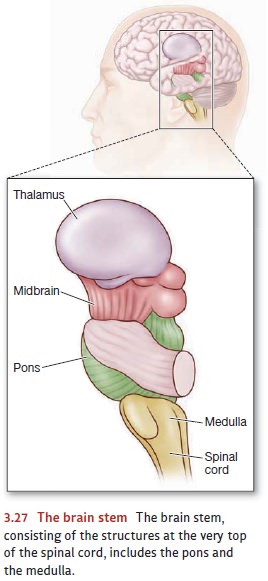
cream cone. Structures at the very top of the cord—looking roughly like the ice
cream on top of the cone—form the brain
stem (Figure 3.27). The medulla is
at the bottom of the brain stem; among its other roles, the medulla controls
our breathing and blood circulation. It also helps us maintain our balance by
controlling head orientation and limb positions in relation to gravity. Above
the medulla is the pons, which is one
of the most
important brain areas for
controlling the brain’s overall level of attentiveness and helps govern the
timing of sleep and dreaming.
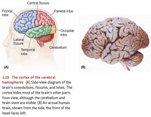
Just behind the brain stem is the
cerebellum (Figure 3.28A). For many
years, inves-tigators believed the cerebellum’s main role was to control
balance and coordinate movements—especially rapid and carefully timed
movements. Recent studies confirm this role, but suggest that the cerebellum
also has a diverse set of other functions. Damage to this organ can cause
problems in spatial reasoning, in discriminating sounds, and in integrating the
input received from various sensory systems (J. Bower & Parsons, 2003).
Sitting on top of the pons are
two more structures—the midbrain and thalamus (see Figure 3.27). Both of
these structures serve as relay stations directing information to the
forebrain, where the information is more fully processed and interpreted. But
these structures also have other roles. The midbrain, for example, helps
regulate our experience of pain and plays a key role in modulating our mood as
well as shaping our motivation.
On top of these structures is the
forebrain—by far the largest part of
the human brain. Indeed, photographs of the brain (Figure 3.28B) show little
other than the forebrain because this structure is large enough in humans to
surround most of the other brain parts and hide them from view. Of course, we
can see only the outer sur-face of the forebrain in such pictures; this is the cerebral cortex (cortex is the Latin word for “tree bark”).
The cortex is just a thin
covering on the outer surface of the brain; on average, it is a mere 3 mm
thick. Nonetheless, there is a great deal of cortical tissue; by some
estimates, the cortex constitutes 80% of the human brain (Kolb & Whishaw,
2009). This consider-able volume is made possible by the fact that the cortex,
thin as it is, consists of a very large sheet of tissue; if stretched out flat,
it would cover roughly 2.7 square feet (2,500 cm2). But the cortex
isn’t stretched flat; instead, it’s crumpled up and jammed into the limited
space inside the skull. This crumpling produces the brain’s most obvious visual
feature—the wrinkles, or convolutions, that cover the brain’s outer surface.
Some of the “valleys” in between
the wrinkles are actually deep grooves that divide the brain into different
anatomical sections. The deepest groove is the longitudinal fissure, run-ning from the front of the brain to the
back and dividing the brain into two halves— specifically, the left and the
right cerebral hemispheres. Other
fissures divide the cortex in each hemisphere into four lobes, named after the
bones that cover them—bones that, as a group, make up the skull. The frontal lobes form the front of the
brain, right behind the forehead. The central
fissure divides the frontal lobes on each side of the brain from the parietal lobes, the brain’s topmost
part. The bottom edge of the frontal lobes is marked bythe lateral fissure, and below this are the temporal lobes. Finally, at the very back of the brain—directly
adjoining the parietal and temporal lobes—are the occipital lobes.
As we’ll see, the cortex—the
outer surface of all four lobes—controls many func-tions. Because these
functions are so important to what we think, feel, and do, we’ll address them
in detail in a later section. But the structures beneath the cortex are just as
important. One of these is the hypothalamus,
positioned directly under-neath the thalamus and crucially involved in the
control of motivated behaviors such as eating , drinking , and sexual activity
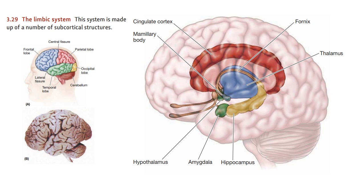
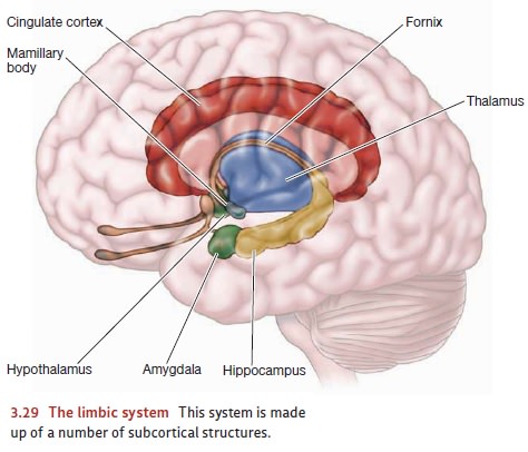
. Surrounding the thal-amus and hypothalamus is a set of interconnected
structures that form the limbicsystem (Figure
3.29). This system—especially one of its parts, the amygdala—plays a key role in modulating our emotional reactions
and seems to serve roughly as an “evaluator ” that helps determine whether a stimulus
is a threat or not, famil-iar or not, and so on. Nearby is the hippocampus, which is pivotal for
learning and memory as well as for our navigation through space. (In some
texts, the hippocam-pus is considered part of the limbic system, following an
organization scheme laid down more than 50 years ago. More recent analyses,
however, indicate that this ear-lier scheme is anatomically and functionally
misleading. For the original scheme, see MacLean, 1949, 1952; for a more recent
perspective, see Kotter & Meyer, 1992; LeDoux, 1996.)
Related Topics