Chapter: Psychology: The Brain and the Nervous System
Methods for Studying the Nervous System: Studying the Effects of Brain Damage

Studying the
Effects of Brain Damage
In many cases, we gain further
insights into brain functioning by asking what happens if the brain is somehow changed—either deliberately in our
research or through accident or injury. For example, in the laboratory, we can
use chemicals or weak applications of electricity to stimulate particular brain
areas and then observe the resulting changes in behavior. Likewise, we can use
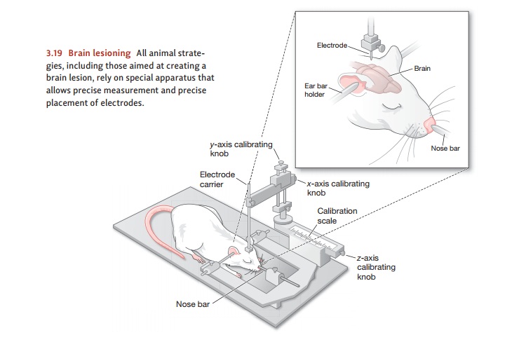
different chemicals, or slightly stronger electric currents, to create a brain lesion—damage to the brain cells—at a particular site and then com-pare
how the brain functions with this damage to how it functions when the site is
unharmed. Alternatively, we can disrupt the flow of information into or out of
an area by cutting—technically, transecting—the relevant pathways and then
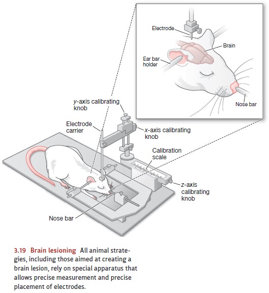
observing the result (Figure 3.19).
Although each of these techniques
has been a valuable source of new knowledge, it should be obvious that these
procedures, which involve altering the brain of another liv-ing creature, raise
some ethical questions. In some cases, the ethical concerns can be weighed
against the direct medical benefits of a procedure. (Lesions or transections
are sometimes used to treat extreme cases of epilepsy; even though these
procedures are car-ried out for therapeutic purposes, we can still gain
valuable information by studying the results.) In other cases, the brain
manipulation is done not for medical treatment but as

part of a scientific study; here,
the ethical concerns must be weighed against the study’s potential scientific
benefits. These benefits are, however, often large; for example, stud-ies of
brain lesions have allowed enormous progress in our understanding of
Parkinson’s disease, multiple sclerosis, and Alzheimer’s disease. This progress
will undoubtedly con-tribute to new forms of treatment for these conditions.
NEURO PSYCHOLOGICAL STUDIES
Outside of the laboratory, people
sometimes suffer disease or injuries that can cause damage to the brain. These
naturally occurring cases are tragic, but they can provide information useful
both for researchers and for those involved in treating the afflicted
individual. The study of these cases defines the special field of neuropsychology—the effort to gain insights into the brain’s function by closely
examining individuals who have suffered some form of brain damage.
Early studies in neuropsychology
relied on clinical observation of patients with brain damage or disease: What
were their symptoms? How was their behavior disrupted? For example, consider
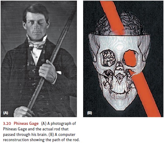
the grisly case of Phineas Gage, who in 1848 was working as a con-struction
foreman. Gage was preparing a site for demolition when some blasting powder
misfired and launched a 3-foot iron rod into his cheek, through the front part
of his brain, and out the top of his head (Figure 3.20). Gage lived, but not
well. As we’ll discuss later, he suffered intellectual and emotional
impairments that gave researchers valuable clues about the roles of the brain’s
frontal lobes (Valenstein, 1986).

In Gage’s case, the brain damage was clear; the task for investigators was to define the consequences of the damage. In other cases, the situation is reversed: The behavioral effects are obvious, but the nature of the brain damage needs to be discovered. More than a century ago, physicians were able to describe the disruption itself—the difficulties their patients had in speaking or understanding. It was not until years later that they identified the affected brain regions—regions that we now know are crucial for language.
In more recent studies,
neuropsychologists have moved beyond clinical observation. Now they often do
fine-grained experiments to find out exactly what a brain-damaged individual
can and cannot do. These experiments are usually combined with the
neu-roimaging techniques we’ll describe in a moment, so that the investigators can
match a precise picture of the person’s brain damage with an equally precise
assessment of her deficits.
TRANSCRANIAL MAGNETIC STIMULATIONSTUDIES
Over the past 25 years,
investigators have developed a new experimental technique that can produce temporary brain disruption and allow
investigators to perform experiments that would otherwise not be possible. This
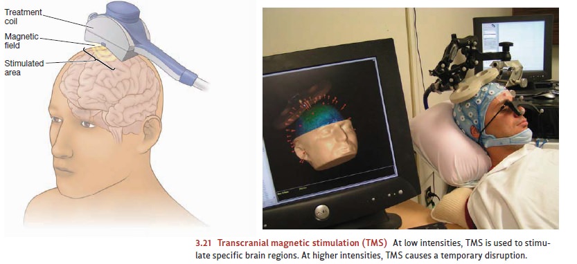
technique—transcranial magnetic
stimula-tion (TMS)—involves creating a series of strong magnetic pulses at
a particular locationon the scalp (Figure 3.21). At certain intensities and
frequencies, these pulses stimulate
the brain region directly underneath that scalp area; thus, investigators can
use TMS to activate otherwise sluggish brain regions.

At other intensities, though, TMS
has a different effect: It creates a temporary dis-ruption in the brain region
just beneath the scalp—allowing investigators to “turn off ” certain brain
mechanisms for a brief period of time and observe the results (Fitzgerald,
Fountain, & Daskalakis, 2006). This technique obviously presents an
enormous range of experimental options and lets investigators examine in detail
the function of this or that brain region. However, TMS does have limits as a
research tool: It influences only structures near the surface of the brain
(i.e., immediately inside the skull). Even so, TMS is a powerful addition to
the neuroscientists’ tool kit.
We should also mention that TMS is more than just a research tool, because investigators are also exploring its therapeutic potential (e.g., Gershon, Dannon, & Grunhaus, 2003). Some uses are already in place: In late 2008, the U.S. Food and Drug Administration approved the use of TMS as a therapeutic procedure for some cases of clinical depression.
Related Topics