Chapter: Psychology: The Brain and the Nervous System
Activity and Communication within the Neuron

Activity and
Communication within the Neuron
The neuron is, in many respects,
just a cell. It has a nucleus on the inside and a cell membrane that defines
its perimeter. In the middle is a biochemical stew of ions, amino acids,
proteins, and DNA along with a collection of smaller structures that provide
for the metabolic needs of the cell itself. What makes a neuron distinctive,
though, is the peculiarity of its cell membrane. The membrane is highly
sensitive to stimulation. Poke it, stimulate it electrically or chemically, and
the neuronal membrane actually changes its structure, producing a cascade of
changes that can eventually lead to an electrical signal called an action potential. This signal—sent from
one end of the neuron to the other—is the neuron’s main response to input as
well as the fundamental information carrier of the nervous system.

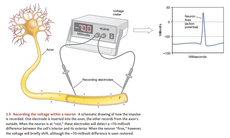
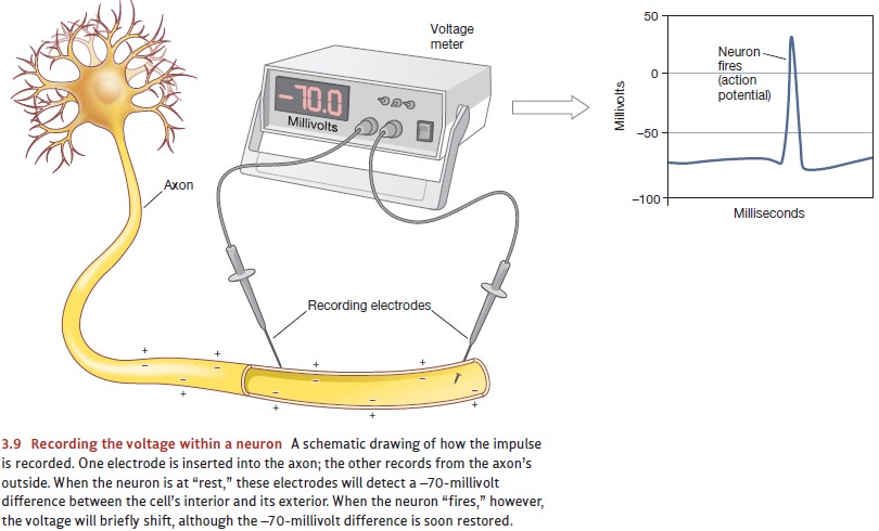
At its heart, the action
potential involves electrical changes. To study these changes, we can begin by
inserting one microelectrode into a neuron’s axon and placing a second
electrode on the axon’s outer surface (Figure 3.9). This setup allows us to
measure elec-trical activity near the cell’s membrane. It also tells us that
even when the neuron is not being stimulated in any way, there’s a voltage
difference between the inside and the out-side of the cell. Like a miniature
battery with a positive and a negative connection, the inside of the axon is
electrically negative relative to the outside, with a difference of –70 or so
millivolts (a standard AA battery, at 1.5 volts, has over 200 times this
voltage). Because this small voltage difference occurs when the neuron is
stable, it’s traditionally called the neuron’s resting potential.
How does this situation change
when the neuron is stimulated? To find out, we can use a third microelectrode
to apply a brief electrical pulse to the outside surface of the cell. This
pulse reduces the voltage difference across the membrane. If the pulse is weak,
it may reduce the voltage difference a little bit, but the neuron’s mem-brane
will maintain its integrity and quickly work to restore the resting potential
of –70 millivolts. But if the pulse is strong enough to push the voltage
difference past a critical excitation
threshold (about –55 millivolts in mammals), something dramatic happens. In
that region of the cell, the voltage difference between the inside and outside
of the membrane abruptly collapses to zero and, in fact, begins to reverse
itself. The inside of the membrane no longer shows a negative voltage compared
to the outside; instead, it suddenly swings positive—up to +40 millivolts. This
momen-tary change in voltage, the action potential, is crucial for the
functioning of the neuron. This change is short-lived, and the resting
potential is restored within a millisecond or so.
Related Topics