Chapter: The Massage Connection ANATOMY AND PHYSIOLOGY : Nervous System
The Brain and Brain Divisions
The Brain and Brain Divisions
The brain, similar to the spinal cord, consists of neu-rons and neuroglia. As a result of the larger number of neurons and greater, complex interconnections, the brain can do a variety of things, with the ability to alter responses according to past experiences. The brain contains 98% of the body’s neural tissue and weighs about 1.4 kg (3 lb).
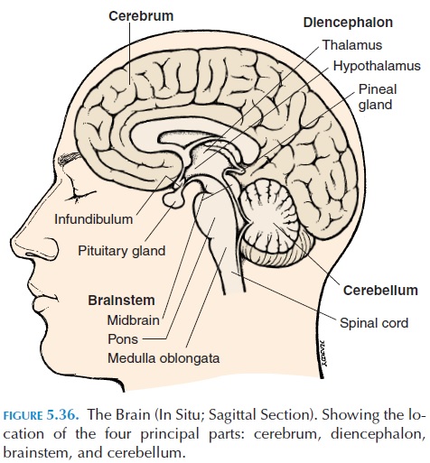
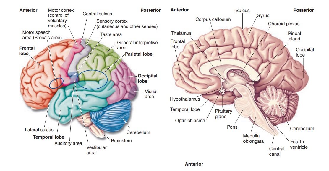
The brain is the upper, enlarged end of the spinal cord. It consists of four principal parts: the cere-brum, the cerebellum, thediencephalon, and the brainstem (Figure 5.36). The brainstem connects thespinal cord to the other principal structures and is di-vided into three parts (from inferior to superior): the medulla oblongata, the pons, and the midbrain(Figures 5.36 and 5.37). Superior to the brainstem is the diencephalon.
The thalamus and the hypothala-mus are important components of the diencephalon. The cerebrum lies over and around the diencephalon and can be divided into two large, cerebral hemi-spheres. Located posterior and inferior to the cere-brum is another enlargement called the cerebellum.
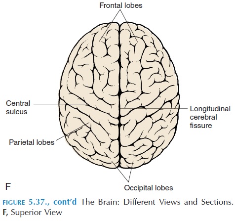
The two cerebral hemispheres are separated into right and left hemispheres by a long, deep depression known as the longitudinal fissure, and the hemi-spheres are connected to each other by the corpuscallosum, which contains nerves that run acrossfrom one hemisphere to the other.
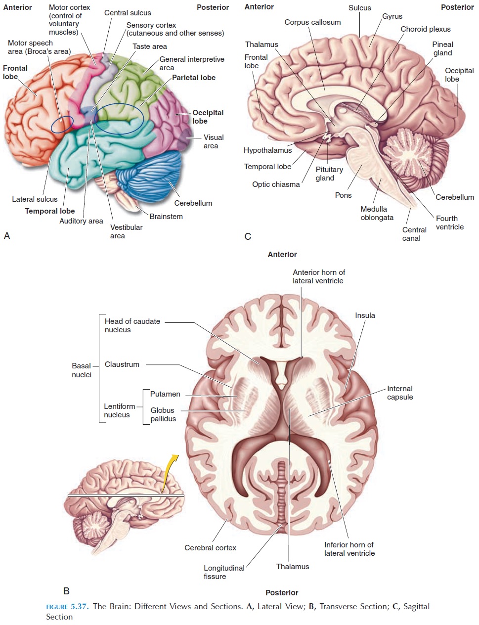
The brain is divided into many lobes named by the skull bone they underlie. The boundaries of the lobes are also determined by specific sulci. The frontallobe is separated from the parietal lobe by the cen-tral sulcus. The lateral sulcus, which runs some-what transversely, separates the frontal from the tem-poral lobe.The occipital lobe, located posteriorly, isseparated from the parietal lobe by the parieto-occipital sulcus. Each lobe can be considered tohave somewhat specific functions; however, the brain is too complex and the activities of the brain too in-terlinked for functional boundaries to be drawn be-tween lobes. The locations of areas that have specific functions are described briefly below.
The gyrus, located posterior to the central sulcus, is called the postcentral gyrus. This is the region where sensory impulses are relayed and is also known as the primary sensory cortex. The sensations of vision,hearing, taste, and smell relay to other areas of the cor-tex. Vision sensations reach the occipital lobe (primaryvisual area), and the auditory and smell sensations arerelayed to the temporal lobe (primary auditory area and primary olfactory area). The taste sensations are relayed to the lateral and inferior part of the postcentral gyrus in the parietal cortex (primary gustatory area). Anterior to the central sulcus is the precentral gyrus, which initiates motor commands to the motor neurons in the brainstem and the spinal cord, and is known as the primary motor cortex. The neurons here are pyra-mid-shaped, and the pathway taken by these fibers to the motor neuron is the pyramidal system.
In addition to the primary cortex, there are asso-ciation areas for motor and sensory function thathelp interpret sensations. If the association areas are damaged, a person will perceive the sensation, but will not be able to understand it. For example, if the visual association area is damaged, the person may be able to see a picture of a cup but not understand what it is. Motor association areas are needed to re-lay the proper instructions to the primary motor cor-tex, to bring about smooth, coordinated movements in the right sequence. The details of the control of posture and movement are discussed later.
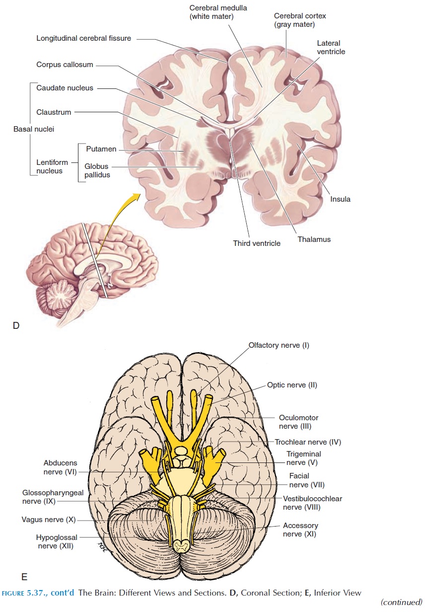
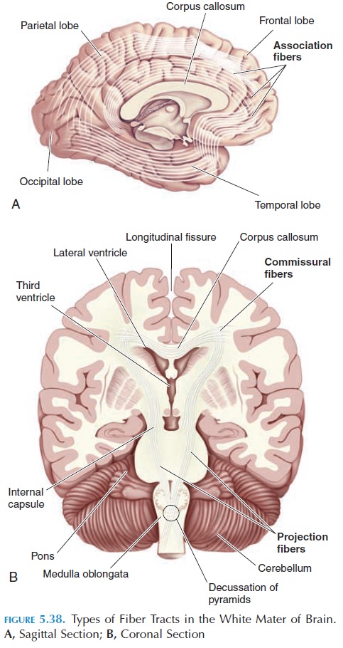
The white mater of the brain contains three differ-ent types of fiber tracts (see Figure 5.38). Some axons interconnect areas of the cerebral cortex in the same hemisphere and are known as association fibers. Others, the commissural fibers, interconnect the two hemispheres; others run between the cortex and other structures such as the cerebellum, brainstem, and spinal cord and are known asprojection fibers. Some important projection fibers that bring sensory impulses to and take motor impulses from the brain run close together in the internal capsule region. Because of the close proximity of fibers to and from different parts of the body in this region, a blockage to blood flow in the internal capsule can cause exten-sive damage to the opposite side of the body.
INTEGRATIVE CENTERS
Prefrontal Lobe
The brain also has areas that integrate and process various sensory information and then perform com-plicated and complex motor activities and analytic functions (Figure 5.37). The prefrontal areas inte-grate information from sensory association areas and perform various intellectual functions. Based on past experience, this area is able to predict the consequences of different responses. Frustration, anxiety, and tension may be generated.
General Interpretive Area or Wernicke’s Area
This area, usually located on the left hemisphere (Fig-ure 5.37A), is important in integrating visual and au-ditory memory. Injury to this area affects the ability to understand and interpret what is seen or heard. Indi-vidual words may be understood but, when words are put together, the meaning may not be interpreted.
Speech Center
This center, the Broca’s speech area, is located near the Wernicke’s area, in the same hemisphere along the precentral gyrus. This center regulates respira-tion and the various muscles required for speech.
RIGHT AND LEFT HEMISPHERESPECIALIZATIONS
Left hemisphere.In most people, the general inter-pretive areas and the speech centers exist in the left hemisphere. Therefore, this hemisphere is responsi-ble for skills related to language, such as reading, writing, and speaking. Analytical tasks are also per-formed here. Hence, the left hemisphere is known as the categorical hemisphere.
Right hemisphere.This hemisphere helps analyzesensory information, such as facial recognition, emo-tion interpretation, music, art, and smell differentiation and is known as the representational hemisphere.
The distribution of the described functions varies individually; however, in the majority of both right-and left-handed individuals, the left hemisphere is the categorical hemisphere.
THE LIMBIC SYSTEM
The limbic system (see Figure 5.39) is a collection of nuclei and tracts involved with creation of emotions, sexual behavior, fear, rage, motivation, and processing
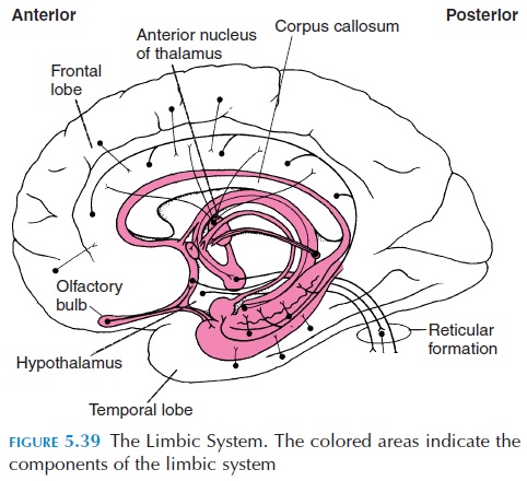
These components of the limbic system are primarily located as a border at the point where the cerebrum is connected to the midbrain. This in-cludes the rim of gyri around the corpus callosum, some parts of the temporal lobe, hypothalamus, thal-amus, and olfactory bulbs, among other regions.
Emotions
Emotions have both physical and mental compo-nents; for example, it requires awareness of the sen-sation and its meaning; the association with past memories; and the urge to act on the emotions and the physical changes that accompany it, such as in-creased heart rate and blood pressure. Hence, the limbic system is complex and has connections with many areas, such as the thalamus (sensory relay sta-tion) and hypothalamus (which links emotions to the autonomic and endocrine system). The paucity of connections with the cortex, which is responsible for voluntary control, is the reason for the inability to turn emotions on and off.
Sexual Behavior
The limbic system, along with the hypothalamus, is responsible for sexual behavior. Although copulation is a result of various reflexes integrated in the spinal cord and brainstem, the behavior that accompanies it is regulated by the limbic system and hypothalamus. In humans, however, it is further conditioned by so-cial and psychic factors.
Fear and Rage
Fear and rage are emotions regulated by the limbic system and hypothalamus. The physiologic changes that accompany them, such as pupillary dilation, cowering, and sweating, are caused by the autonomic system; however, the limbic system is required for initiating the response. Both fear and rage are pro-tective responses to threats in the environment. Again, these emotions are conditioned by social fac-tors and sex hormones.
Motivation
Experiments in which electrodes have been im-planted in certain areas of the brains of animals and humans, with the ability of the experimental ani-mals/humans to stimulate the area using these elec-trodes, have produced interesting findings. If the electrode is implanted in certain areas, pleasure is produced, and the animals/humans tends to stimu-late the area repetitively and continuously. Other ar-eas have been identified that, on stimulation, produce emotions such as fear and terror. These experiments have shown that the body has reward or approach systems and punishment or avoidance systems, which are part of the limbic system and play an im-portant role in motivation.
The neurotransmitters secreted in this system have been identified, and drugs that act on the receptors, production/destruction of these neurotransmitters have an effect on mood and emotion.
THE THALAMUS
The thalamus is a paired structure, located superior to the brainstem on the sides of the third ventricle (Figure 5.37B, C, and D). It is a collection of many nuclei. The thalamus is the principal relay point for sensory information that comes from the spinal cord, brainstem, cerebellum, and parts of the cerebrum. From here, the information is relayed to the sensory cortex. The thalamus does perceive some crude sen-sations of pain, temperature, and pressure (i.e., in the absence of the cerebral cortex, it is possible to per-ceive some crude sensations but the cerebral cortex is needed for proper perception).
The thalamus consists of four major groups of nu-clei. The anterior is connected to the limbic system and deals with emotions. Themedial nuclei provide a conscious awareness of emotional states. The ven-tral nuclei relay sensory information from the rest of the body to the cortex and also monitor communica-tion between the motor cortex and association areas. The posterior nuclei relay sensory information from the eye and ear to the cortex.
In addition to these functions, the thalamus is needed for acquisition of knowledge (cognition) and memory and for planning of movement. (Detail of its other connections and functions are beyond the scope of this book).
THE HYPOTHALAMUS
The hypothalamus (Figures 5.36 and 5.37C, D) is lo-cated close to the thalamus and lies just above the pi-tuitary gland. It also has many collections of neu-rons—nuclei. It is closely associated with the limbic system. In addition, the hypothalamus has important regulatory functions:
· It controls skeletal muscle contractions that ac-company various emotions, such as changes in facial expression and muscle tone.
· It coordinates the activities of the centers that control respiration, heart rate, blood pressure, and digestion that are located in the pons and medulla.
· It secretes various stimulatory and inhibitory hormones into the blood, which are trans-ported to the pituitary gland located close to the hypothalamus, where they regulate pitu-itary hormone secretion (Figure 6.4). Certain neurons located in the hypothalamus manufacture antidiuretic hormone and oxy-tocin in their cell bodies. Axons from these cell bodies project into the posterior pituitary and secrete the hormones into the blood in vessels perfusing this region.
· It has centers, such as feeding centers and thirst centers, that modify emotions and behavior to fulfill such basic needs. Neurons located in the hypothalamus monitor blood glucose levels and blood volume and alter behavior accordingly.
· It has numerous connections with the rest of the brain and is a link between voluntary, endocrine, and autonomic functions. It controls and inte-grates the activities of the autonomic nervous system. For example, even the thought of stress-ful situations increases heart rate and blood pressure and produces many other physiologic changes similar to the fight-or-flight reaction.
· It has certain nuclei responsible for maintain-ing body temperature. Neurons located here monitor the core body temperature and pro-duce such changes as sweating, shivering, and vasodilation or constriction in cutaneous blood vessels by regulating the vasomotor centers and other centers located in the medulla and pons.
· It plays a major role in the establishment of sleep patterns.
THE BRAINSTEM: MIDBRAIN,PONS, AND MEDULLA
Together these three regions are known as the brain-stem (Figure 5.37A, C, E). They lie between the brainand the spinal cord, with the midbrain closer to the cerebrum and the medulla closer to the spinal cord. The brainstem contains gray (nuclei) and white mater (tracts). Collections of neurons (nuclei) located here control basic vegetative functions such as heart rate, blood pressure, and respiratory rate. Other nu-clei give rise to the various cranial nerves. Numerous ascending and descending tracts synapse and/or pass through the brainstem.
Nuclei located in the midbrain coordinate muscles in relation to received visual and hearing input. For ex-ample, movement of the eyeballs and turning of the head, neck, and trunk as one follows an object or hears a loud noise are regulated here. Midbrain nuclei that receive input from the vestibular apparatus and other sensory areas help the body to alter the posture to maintain balance.
Related Topics