Chapter: The Massage Connection ANATOMY AND PHYSIOLOGY : Nervous System
Muscle Spindle - Nervous System
MUSCLE SPINDLE
Knowledge of the structure and function of the muscle spindle is key to the understanding of muscletone. Muscle tone is the normal resistance of the muscle to stretch. The absence or marked increase or decrease in muscle tone in the various conditions a bodyworker encounters is a result of variations in the activity of the muscle spindle.
Structure of Muscle Spindle
In brief, each muscle spindle consists of 2–10 muscle fibers enclosed in a connective tissue capsule. The muscle fibers in the spindle are known as intrafusal fibers, to distinguish them from the regular muscle fibers that produce contraction, the extrafusal fibers. The intrafusal fibers are rather im-mature, with fewer striations. The striations or con-tractile units are located more toward the two ends of the spindle, with the nuclei located near the middle. The intrafusal fibers are positioned in parallel with the rest of the muscle fibers, and the ends of the cap-sule are attached to the tendon of the muscle on ei-ther side.
Sensory Nerves From the Muscle Spindle
There are two different sensory nerve endings. Some nerve endings wind around the center of the spindle, while others branch delicately on either end. These sensory neurons synapse directly with the motor neu-ron to the same muscle.
Motor Nerves to the Muscle Spindle
In addition to the sensory nerves that leave it, muscle spindles have motor nerves that innervate the intra-fusal muscle fibers. These motor nerves are impor-tant and actually constitute 30% of the fibers in the ventral root. These motor nerves are the gamma ef-ferents or gamma motor neurons, and the motornerves to the extrafusal fibers are known as alpha ef-ferents or alpha motor neurons.
Function of the Muscle Spindle
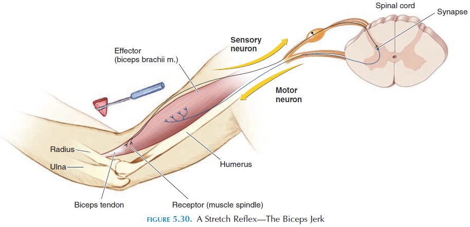
When the muscle spindle is stretched, the mechanical stimulus is converted to action potentials that travel, via the sensory nerve, directly to the motor neuron supplying the muscle, causing it to contract (Figure 5.30). If a muscle is stretched, the muscle spindle is also stretched as it lies parallel to the muscle fibers and a stretch reflex is elicited. If the muscle contracts, nothing happens because the spindle is also short-ened. In this way, the spindle and its reflex connections help maintain muscle length. The muscle spindle is also able to detect changes in the rate of stretch and al-ter the frequency of action potentials to the extrafusal muscle fibers accordingly, to cause a smooth contrac-tion. If muscle spindles were not present, contraction of the muscle would be jerky and tremors will be seen.
Function of the Gamma Motor Nerve
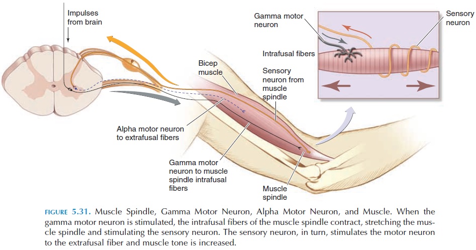
It was mentioned that the gamma motor neurons in-nervate the intrafusal fibers and that the contractile units of the intrafusal fibers are located toward the ends of the spindle (see Figure 5.31). If the gamma motor neuron is stimulated, it causes the intrafusal fibers to contract. Contraction of these fibers at both ends of the spindle results in stretching of the middle region of the spindle where the sensory nerves are lo-cated. The stretch is detected by the sensory nerve endings, and they produce action potentials that cause the extrafusal fibers to fire. How is this impor-tant? The nervous system, by stimulating gamma mo-tor neurons, can make the muscle more or less sensi-tive to stretch. The gamma motor neurons are responsible for muscle tone.
Control of Gamma Motor Neuron Discharge
The gamma motor neurons are regulated by descending tracts from certain areas of the brain. Via these neurons, the sensitivity of the muscle spindles and, hence, the stretch reflexes and muscle tone can be al
Many factors af-fect the gamma motor neuron discharge; for example, anxiety and stress increase its discharge, resulting in tensing of muscles and hyperactive tendon reflexes. If the skin of the hand on one side is stimulated by a painful stimulus, it results in increased discharge to the flexors and decreased discharge to the extensors of the same side, facilitating flexion and quick re-moval. At the same time, the opposite happens in the other side, adjusting posture and weight distribution.
Related Topics