Chapter: 11th 12th std standard Class Physics sciense Higher secondary school College Notes
Electron microscope: Construction, working, Uses, Limitations
Electron microscope
The electron microscope, like optical
microscope, is an instrument principally used in the research laboratory for
magnifying small objects. The wave nature of moving electron is the basis for
the electron microscope. The resolving power of a microscope is the least
distance between two points which can be distinguished. The resolving power of
a microscope is limited by the wave length of the radiation used. In optical
microscope, the visible light is used to illuminate the object and the highest
magnification obtained with the best optical microscope is about 2000. Since,
the wavelength of X-rays is smaller than that of the visible light, one can
think of having an X-ray microscope. However, X-rays cannot be focussed as
visible radiations are focussed using lenses. On the other hand, electrons
having de Broglie wavelength of the order of X-rays can be focussed easily
using electric and magnetic fields and one can build a high resolving power
microscope using electrons.
For electrons accelerated by a potential difference of about 60,000
volts, the wavelength is about 5 � 10- 12 m. This is 105
times smaller than that of visible light. Hence the resolving power of an
electron microscope will be 1,00,000 times greater than that of an optical
microscope.
Construction and working
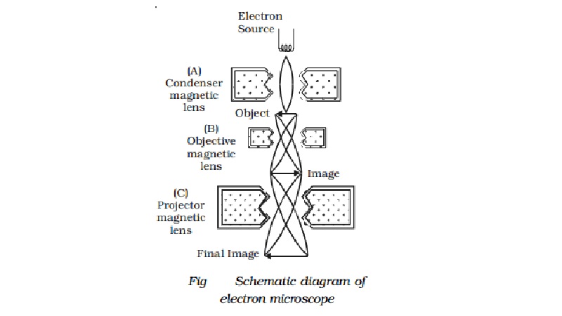
The schematic diagram of an electron microscope is shown in Fig. An
electron microscope is similar in principle to an optical microscope. The
modern electron microscope is usually of transmission type in which magnetic lenses
of short focal length are used to obtain large magnification. An electron beam
emitted by a filament is accelerated through a large potential difference in a
device called electron gun. The fine beam of electrons is made to pass through
the centre of the doughnut shaped electromagnet A (condenser magnetic lens).
The electrons get deflected to form a parallel beam which strikes the object to
be magnified. It should be noted that the electrons will be transmitted more
through the transparent parts of the object and transmitted in less number
through comparatively denser portions. The transmitted beam will thus have the
likeness of the object traversed by it. The second electro magnet B (objective
magnetic lens) causes the electron beam to diverge to produce enlarged image of
the object. The electromagnet C (projector magnetic lens) focusses the electron
beam from the part of the enlarged image on the fluorescent screen producing
still greater magnification. The image obtained on the fluorescent screen is
made visible by scintillation for direct view. It can also be obtained on a
suitable photographic plate for a permanent record. Sharp focussing is obtained
by adjusting the intensity of magnetic fields produced by electro magnets.
Since, the electron beam operates in vacuum, the apparatus is mounted in a
chamber which is completely evacuated.
Uses:
1.
It is used in the industry, to study the
structure of textile fibres, surface of metals, composition of paints etc.
2.
In
medicine and biology,
it is used
to study virus,
and
3.
bacteria.
4.
In Physics, it has been used in the
investigation of atomic structure and structure of crystals in detail.
Limitations
An electron microscope is operated only in high vacuum. This
prohibits the use of the microscope to study living organisms which would
evaporate and disintegrate under such conditions.
Related Topics