Chapter: The Massage Connection ANATOMY AND PHYSIOLOGY : Cardiovascular System
Venules and Veins - Structure And Function of Blood Vessels
Venules and Veins
The capillaries empty into small veins known as venules. In some areas, there are direct connectionsbetween arterioles and venules. This is referred to as arteriovenous anastomoses (AV anastomoses) or shunts. Such anastomoses allow blood to bypass thecapillaries and flow directly into veins. Shunts are in abundance in the skin and are controlled by the sym-pathetic nervous system. They permit the blood flow to be varied over a wider range —a useful function for temperature regulation.
The venules form thin-walled, midsized veins that join and rejoin to form large veins. The branches of veins are referred as tributaries. In most regions, the veins accompany arteries and lie parallel and close to them.
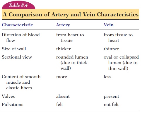
The walls of the veins have three layers; how-ever, they are much thinner than those of arteries. The lumen of the vein is much larger than that of ar-teries and can, therefore, accommodate more blood than arteries. Hence, the veins may serve as blood reservoirs. Many veins also have valves in them. Ve-nous valves are thin folds of the tunica interna that direct blood toward the heart. Valves help with the venous return by preventing backflow of blood.
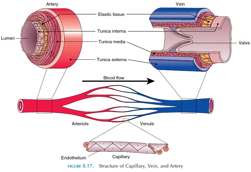
In some regions, the vein is very thin, with just a thin endothelial wall and no smooth muscles. The surrounding connective tissue replaces the tunica media and externa. Such veins are known as venoussinus or vascular sinus. Venous sinus are found inthe cranial cavity—between the dura mater. The coronary sinus of the heart (the vein that drains the wall of the heart) is also a vascular sinus.
Venous Return
As blood flows from larger arteries through arterioles and capillaries, the blood pressure drops and is only 10% of that in the arteries when it reaches the venules. The pressure is so low that it cannot even op-pose the force of gravity. The volume of blood return-ing to the heart from the veins—the venous return— depends on the pressure difference between the venules to the right ventricle. Although the pressure difference is low, venous return is equal to the amount of blood pumped out of the heart as a result of additional mechanisms.
To help with the venous return, veins have one-way valves, which are actually folds of tunica interna. These folds project into the lumen, allowing blood to flow only toward the heart. In addition to valves, the pulsation of the arteries lying parallel to them and compression resulting from contracting skeletal mus-cle (skeletal muscle pump) that surrounds them helps to squeeze venous blood towards the heart against the force of gravity.
Respiratory movement also helps to return blood to the heart (respiratory pump). During inspiration, pressure decreases in the thoracic cavity and the tho-racic volume increases. At the same time, the pres-sure increases in the abdominal cavity as the di- aphragm descends during inspiration. This squeezes the abdominal veins. The resultant unequal pressure creates a “sucking” effect that pulls blood toward the heart.
Related Topics