Chapter: The Massage Connection ANATOMY AND PHYSIOLOGY : Cardiovascular System
Systemic Veins
SYSTEMIC VEINS
Drainage From the Brain, Head, and Neck
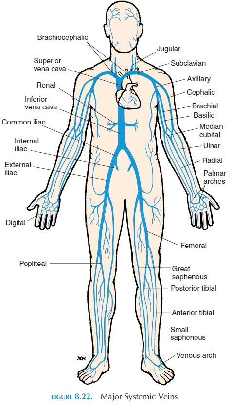
Figure 8.22 is an overview of the major veins. The superior vena cava receives blood from the tissue and organs of the head, neck, chest, shoulders, and upper limb. Inside the skull, the smaller veins drain into large vessels known as sinus. The venous sinus con-verge and ultimately leave the skull as the internaljugular vein. The internal jugular vein descends par-allel to the common carotid artery in the neck. Poste-riorly, the skull is drained by the vertebral veins that leave the skull and descend within the foramen in the transverse processes of the cervical vertebrae.

From regions outside the skull and neck, the veins of the head drain into the external jugular vein,which lies just beneath the skin, on the anterior sur-face of the sternocleidomastoid muscle.
Drainage From the Upper Limbs
The upper limb, like the lower limb, has two sets of veins—the superficial and the deep. In the hand, dig-ital veins empty into the superficial and deep pal-mar veinsthat interconnect to form the palmar ve-nous arches (similar to the palmar arches of arteries).
The superficial veins empty into the cephalic vein, which ascends along the radial side of the forearm, the median antebrachial vein, and the basilic vein, which ascends on the ulnar side. The basilic vein as-cends in the upper arm, medial to the biceps brachii muscle. Anterior to the elbow, the superficial mediancubital vein passes medially and obliquely from thecephalic to the basilic vein. Usually, blood is collected for examination or donation from the median cubital vein.
The deep veins are positioned parallel to the arter-ies and carry the same names. The deep palmarveins drain into the radial vein and ulnar vein. Af-ter crossing the elbow, these veins fuse to form the brachial vein. The brachial vein, on entering the ax-illa, is called the axillary vein. The superficial basilic vein joins the brachial vein near the axilla.
The cephalic vein joins the axillary vein on the lat-eral surface of the first rib to form the subclavianvein. The subclavian passes medially and mergeswith the internal and external jugular veins on each side to form the brachiocephalic vein. At the level of the first and second rib, near the heart, the right and left brachiocephalic veins join to form the superiorvena cava. The superior vena cava is joined by the azygos vein inside the thorax. The azygos vein col-lects blood from the thoracic cavity.
Drainage From the Lower Limbs
The lower limbs and abdomen are drained by the in-ferior vena cava. In the foot, the capillaries in the sole of each foot form plantar veins that join to form the plantarvenous arch. Similar to the upper limb, there are two sets of veins—superficial and deep. The deep veins lie parallel to the arteries and are called by the same names as the arteries. The anterior tibial vein, the posterior tibial vein, and the peroneal veins join at the popliteal fossa to form the popliteal vein. On reaching the femur, the popliteal vein is referred to as the femoral vein. The femoral vein penetrates the ab-dominal wall and becomes the external iliac vein.
The surface anatomy of the superficial veins is im-portant, as it is a common site for varicosities. Some of the capillaries of the foot join on the superior surface of the foot to form the dorsal venous arch. Two su-perficial veins are formed from the dorsal venous arch—the great saphenous vein and the small saphe-nous vein. The great saphenous vein ascends along the medial aspect of the leg and thigh and drains into the femoral vein near the inguinal region. The small saphe-nous vein arises from the dorsal venous arch and as-cends along the posterior and lateral aspect of the calf where it drains into the popliteal vein.
Drainage From the Pelvis and Abdomen
In the pelvis, the external iliac vein is joined by the internal iliac vein on each side, to form the rightand left common iliac veins. At the level of L5, the right and left common iliac veins join to form the in-ferior vena cava.
The inferior vena cava lies posterior to the peri-toneal cavity, parallel to the aorta. Some of the major veins that join it in the abdomen are the lumbarveins, gonadal veins(from the testis/ovaries), he-patic veins (from the liver), renal veins (from thekidney), suprarenal veins (from the adrenal glands), and phrenic veins (from the diaphragm).
Portal Circulation
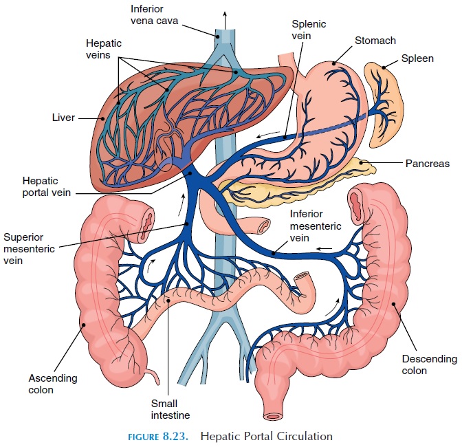
The circulation of blood in the stomach and intestines is unique in that the capillaries formed in the walls of the gut eventually drain into the hepatic portal vein, which empties into sinusoidal capillaries in the liver (see Figure 8.23). The inferior mesenteric, splenic, and superior mesenteric veins are some important tributaries that join to form the hepatic portal vein. This circulation is referred to as the hepatic portalsystem. The blood in the hepatic portal vein containsnutrients absorbed from the gut, which it carries to the liver for storage, metabolic conversion, or excre-tion. After passing through the liver sinusoids, blood collects into the hepatic veins that empty into the in-ferior vena cava. This unique circulation is important because the liver helps maintain the composition of blood in the systemic circulation, even if the digestive activities vary greatly. Simply put, it does not allow glucose or other nutrients to be dumped into the sys-temic circulation every time an individual eats!

The liver not only receives deoxygenated blood from the hepatic portal vein, but also oxygenated blood via the hepatic artery, a branch of the celiac trunk. This blood, together with blood from the por-tal vein, drains into the hepatic veins.
Related Topics