Chapter: The Massage Connection ANATOMY AND PHYSIOLOGY : Cardiovascular System
Cardiac Cycle - The Heart
Cardiac Cycle
Many cyclical or sequential events occur in the heart between the beginning of one heartbeat and the next. All events that occur associated with one heartbeat are known as the cardiac cycle (see Figure 8.15). During the cycle, each heart chamber contracts and relaxes. The period of contraction is known as sys-tole, and the period of relaxation is known as dias-tole. At a heart rate of 75 beats/minute, the durationof each cardiac cycle is about 800 milliseconds, with systole lasting for about 270 milliseconds and dias-tole for about 530 milliseconds. When the heart rate increases, the duration of diastole is shortened more than the systole. This results in the reduction of time available for ventricular filling (occurring when the ventricle relaxes). If the heart rate is unusually fast, there is too little time for the ventricles to fill and lit-tle blood is pushed into the large arteries. The person becomes unconscious as a result of reduced blood flow to the brain.
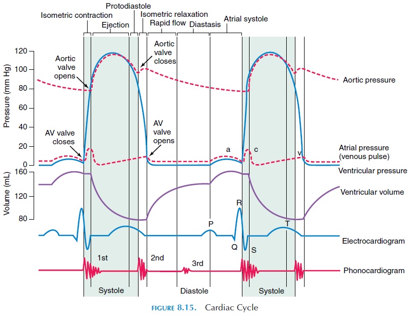
During systole or contraction phase, the blood in-side the chamber is ejected out and, during diastole or relaxation phase, the chamber gets filled with blood and the cycle continues. The right direction of blood flow is determined by the presence of valves and difference in pressure between the chambers.
For proper flow, the sequence of contraction of the atria and ventricle has to be precise, with the atria contracting just before the ventricles for blood to flow from the atria into the ventricles. The presence of the conducting system and the difference in rate of impulse conduction through them ensures that atrial and ventricular muscles contract at different times.
Atrial Diastole and Systole
Blood flows into the atria via the superior and infe-rior vena cavas during atrial diastole. The atrial dias-tole lasts for about 700 milliseconds. When the SA node produces an action potential, the atria contract (atrial systole). Because the impulse takes time to reach the ventricular muscles, the ventricles are re-laxed at this time, and the AV valves are open and blood from the atria enter the ventricle easily.
Ventricular Systole and Diastole
When the impulse reaches the ventricles, they contract (ventricular systole). The pressure in the ventricles rises above that of the pulmonary trunk and aorta, and the semilunar valves that were closed during the dias-tole of the ventricle are pushed open and blood is ejected into the blood vessels. At the same time, the AV valves are pushed close (Figure 8.9). This is primarily responsible for the first heart sound or the “lubb” heard when using the stethoscope or placing the ear or hand on the chest wall superficial to the heart.
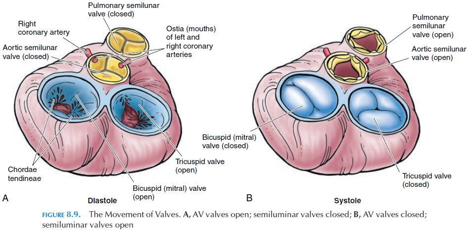
The muscles begin to relax after about 270 millisec-onds (i.e., when the heart rate is 75 beats/minute). When the ventricles relax, the volume increases and the pressure decreases. When the pressure drops be-low that of the pulmonary trunk and aorta, the semi-lunar valves (Figure 8.9) close with a snap. This is re-sponsible for the second heart sound or the “dubb” sound. Because the pressure is less than that of the atria, the AV valves open and blood flows in.
The first and the second heart sounds give an idea of the duration and activities of the cardiac cycle. The duration between the first and second sound indi-cates the duration of ventricular systole; the duration between the second and first sound, the duration of ventricular diastole.
Pressure Changes
The blood pressure in the two atria is just a few mm Hg. The pressure in the right ventricle is much less than that of the left and is equal to about 25 mm Hg. This is because the lungs offer little resistance to blood flow. Because the left ventricle has to pump blood to the rest of the body, it encounters more re-sistance, and the pressure in the left ventricle is as high as 120 mm Hg during systole. The pressure in both the ventricles drops close to 0 mm Hg during the relaxation phase.
The blood pressure in the aorta, while reaching 120 mm Hg during ventricular systole, does not drop to 0 mm Hg during diastole. Instead, the pressure is about 90 mm Hg. This is because the elastic walls of the aorta expand when blood rushes in during systole and recoil back during diastole, increasing the pres-sure in the lumen. For the same reasons, the pressure in the pulmonary trunk is higher than pressure in the right ventricle during diastole.
The pressure in the blood vessels during ventricu-lar systole is known as the systolic pressure and that during diastole is known as the diastolic pressure. It is usually expressed as systolic pressure/diastolic pressure and, in this case, the blood pressure in the aorta is 120/90 mm Hg.
Volume Changes
When the ventricle contracts, not all of the blood is ejected into the large blood vessels. There is some blood remaining in the chamber, and this is known as the end-systolic volume. The volume of blood ejected during each contraction is known as the stroke volume, and it is equal to about 70 mL. Whenthe heart contracts more forcefully, the stroke volume increases and end systolic volume decreases. The vol-ume of blood ejected from the ventricle every minute is known as the cardiac output.In a resting supine man, it averages about 5.0 liters/minute (heart rate stroke volume).
The volume of blood pumped out of the ventricles depends on the volume of blood returning to the heart through the veins. This volume is known as venous re-turn.Venous return is normally about 5.0 liters/minute(5.3 qt/min) and depends on the cardiac output (vol-ume of blood pumped out), blood volume (volume of blood inside the blood vessels), skeletal muscle activity (blood flow through the veins is facilitated by the con-traction and relaxation of the skeletal muscles around it—skeletal muscle pump), and all other factors that af-fect the rate of blood flow through the vena cava.
The volume of blood remaining in the ventricle at the end of ventricular diastole is known as the end-diastolic volume. The end diastolic volume deter-mines, to a large extent, the stroke volume (volume of blood pumped out with each ventricular contraction).
Related Topics