Chapter: Microbiology
Structure and Development of Immune System

STRUCTURE AND DEVELOPMENT OF IMMUNE SYSTEM
Immunity
Introduction
Immune system consists of lymphoid cells (both T and B cells) and lymphoid organs. Cells that make up the immune system are dis-tributed throughout the body. They occur predominantly in lympho re-ticular organs. These include: Bone marrow, thymus, lymph nodes, spleen, mucosa associated lymphoid tissues. The cells occupy the intri-cacies of net works formed by reticular cells and fibers supporting frame work. The cells involved in the immune function are: Lymphocytes, monocytes, macrophages of tissues, endothelial cells, mast cells, baso-phils, eosinophils, neutrophils etc. All cells are derived from pluripo-tent, self renewing stem cells of the bone marrow.
Organs of immune system
Based on the different roles they perform, lymphoid organs can be classified into 1) primary or central lymphoid organs and 2) second-ary or peripheral lymphoid organs. Primary lymphoid organs include 1) Thymus, 2) Bone marrow 3) Bursa of Fabricius in birds. In the primary lymphoid organs, the T and B cells mature into antigen recognizing lymphocytes. Secondary lymphoid organs include Lymph nodes, spleen and mucosa associated lymphoid tissue.
Thymus gland
The thymus gland is a bilobed structure. During fetal develop-ment the size of the thymus gland increases and reaches its maximum at birth. After birth the thymus begins to decrease in size and undergoes atrophy with aging .
Thymus structure:
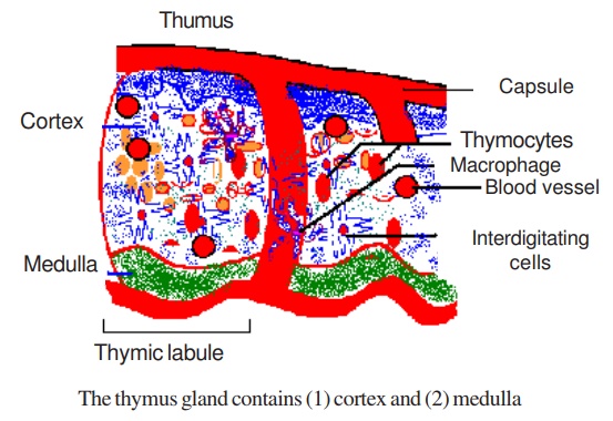
Cortex
The cortex contains epithelial cells. It is densely populated with lymphocytes of various sizes. Most of the lymphocytes in thymus are immature.
Medulla
T lymphocytes mature in the cortex and migrate into the medulla. They leave the medulla and enter the peripheral blood circulation. Then they are transported to the secondary lymphoid organs.
Function of the thymus gland
1. The primary function of the thymus is the production of Thymic lymphocytes
2. Thymus is the major site for lymphocyte proliferation in the body
3. One percent of the lymphocytes produced in the thymus leave the thymus, others are destroyed.
4. lymphocytes produced in the thymus are called T lymphocytes or T cells
5. In the thymus the lymphocytes are educated so that they can pro-duce cell mediated immune (CMI) response against foreign anti-gens
6. Immature lymphocytes mature in the thymus and acquire different Cell differentiation (CD) molecules on their surface
Effects of thymectomy (removal of thymus)
Removal of thymus is called thymectomy. Thymus can be re-moved either in the neonates or in adults.
Neonatal thymectomy results in an immediate severe reduction in the quality and quantity of T lymphocytes.
Adult thymectomy could result in deficiency of T cell, after the eventual death of T cells that originally populated the secondary lym-phoid organs
Thymectomy affects CMI primarily. It also diminishes antibody response to certain antigens
Bone Marrow
Bursa of Fabricius
It is a primary lymphoid organ of birds which is situated near the cloaca. It consists of lymphoid centers that contain epithelial cells and lymphocytes. These lymphocytes are antibody producing cells. The B cells undergo maturation in this organ. Like thymus the bursa is largest at hatching and undergoes atrophy with maturation.
· Mammals do not have Bursa of Fabricius
· A structure in mammals that functions equivalent to that of birds is the bone marrow
Functions of Bursa of Fabricius
1. It produces B lymphocytes. These are responsible for antibody production and Humoral immunity
2. These B cells mature and go to the peripheral lymphoid organs and are seeded there
3. Following appropriate antigenic stimulation, B lymphocytes trans-form into plasma cells and produce antibodies
Secondary Lymphoid Organs
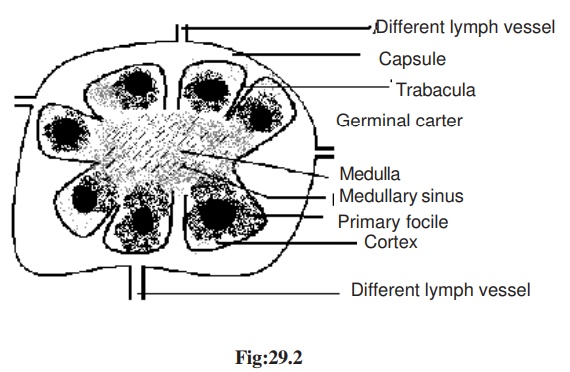
Lymph Node
Lymph nodes are small ovoid structures found in various regions through out the body. Thy form channels called as lymphatic channels through which lymph flows
A Lymph node is surrounded by a fibrous capsule. It has an outer cortex and inner medulla
Cortex
In the cortex the lymphocytes accumulate into primary and sec-ondary follicles. These follicles contain lymphocytes and macrophages![]()
Medulla
In the medulla the lymphocytes are arranged as elongated branch-ing bands or medullary cords. The cortical follicles and medullary cords contain B lymphocytes and constitute bursa/bone marrow dependent areas. Between the cortical follicles and medullary cords, there is a broad, ill-defined intermediate zone called para cortical area. The para cortical area contains T lymphocytes and constitutes the thymus de-pendent area
Functions of lymph node
1. Lymph nodes act as filter for lymph
2. Phagocytosis of foreign particles takes place in lymph node
3. They help the proliferation and circulation of T and B cells
4. They enlarge following local antigenic stimulus
5. The plasma cells secrete antibodies
6. Lymph node is highly efficient in trapping antigen
Spleen
Spleen is the largest of the lymphoid organs. It has a capsule from which descent trabaculae dividing the organ into several intercon-nected compartments. Spleen is made up of white pulp and red pulp
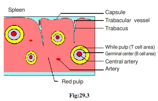
White pulp
The spleenic artery travels along the trabaculae. On entering the spleen the artery divides into arterioles which are surrounded by lym-phoid tissues. This part is called white pulp. White pulp is rich in lym-phoid cells.
Red pulp
It is called red pulp because of the red blood cells. Macroph-ages also are present in this area
Functions of spleen
1. The spleen serves as the grave yard for red blood cells
2. The white pulp is rich in T cells and B cells
3. The T cells are seen peripherally and the B cells are seen centrally
4. After an antigenic stimulus the germinal center containing large num-ber of B cells and plasma cells appear in the periarteriolar areas and almost replace T cells.
Mucosa associated lymphoid tissue
The mucosa lining the alimentary, respiratory, genito-urinary and other surfaces is exposed to antigens. So these areas have rich collec-tion of lymphoid cells
These lymphoid cells are called Peyer’s patches or scattered isolated lymphoid follicles. These are called as Mucosa associated lym-phoid organs (MALT).
Lymphoid tissues in the gut, ie from the adenoids and tonsils up to the follicles of colon are called Gut associated lymphoid tissues (GALT)
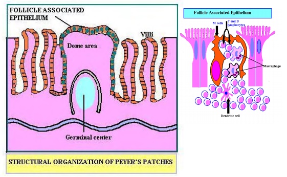
Functions of MALT and GALT
1. They contain lymphoid and phagocytic cells and a special cell called M cells
2. Both B and T cells are present
3. Predominant antibody produced is IgA in the mucosal lining. Oth- ers such as IgG, IgM, and IgE are also produced.
4. Ig A antibodies provide first line of defense against infectious agents
Related Topics