Chapter: 11th Botany : Chapter 2 : Plant Kingdom
Selaginella - Pteridophytes
Selaginella
Division – Lycophyta
Class – Ligulopsida
Order – Selaginellales
Family –Selaginellaceae
Genus – Selaginella
Selaginella
is
commonly called ‘spike moss’. They
are distributed in humid temperate and tropical rain forests. Selaginella rupestris and Selaginella lepidophylla are Xerophytic.
Selaginella
kraussiana, Selaginella chrysocaulos,
Selaginella megaphylla are some
common species. In few Selaginella
species during dry season the entire plant body gets curled and become fresh,
green when moisture is available. Due to this they are called Resurrection plants. Example S. lepidophylla
External morphology
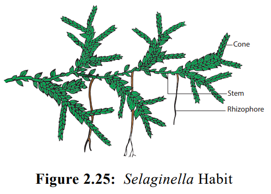
Habit
The plant body of Selaginella is sporophyte (2n) and it is differentiated into root,
stem, and leaves (Figure 2.25). There exist variations in the habit of Selaginella. Some species possess
prostrate creeping system (S. kraussiana);
suberect (S. rupestris); erect (Selaginella erythropus); Climbing (Selaginella alligans). S. oregana is an epiphyte. Most of the
species are perennials. on the basis of structure of stem and arrangement of
leaves, Selaginella is divided into
two sub genera namely Homoeophyllum and Heterophyllum.
Homeophyllum include species with erect stem and spirally arranged leaves. (Example: S. upestris and S. oregana). Heterophyllum include species with prosrate stem with short erect branches and dimorphic leaves (Example: S. kraussiana and S. lepidophylla).
Root
Primary roots are short lived and the adult plant
has adventitious roots. The root may arise at the point of dichotomous
branching or knot like swelling present at the basal portion of the stem. Roots
are endogenous in origin.
Rhizophore
In many species long, cylindrical, unbranched and
leafless structures arise from the lower side of the stem at the point of
dichotomy called rhizophores. They grow vertically downwards and produce tufts
of adventitious roots.
Stem
The stem may be erect, dichotomously branched or
prostrate with lateral branching. The prostrate stem is dorsiventral.
Leaves
The leaves are microphyllous, sessile and simple. A
single midvein is present in the leaves. The vegetative leaf as well as the
sporophyll bears a small membranous tongue like structure on adaxial surface
called ligule. The basal part of the
ligule possess a hemispherical mass of thin walled cells called glossopodium. The function of ligule is
not known, but it is viewed to be associated with water absorption, secretion
and prevent dessication of shoot. The members belonging to Homeophyllum type
possess same type of leaves spirally arranged on the stem whereas the
Heterophyllum type have two types of leaves- two dorsal rows of small
leaves(Microphylls) and two ventral rows of large leaves(Megaphylls).
Internal structure
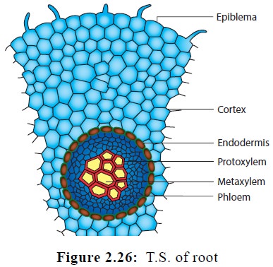
Root
The transverse section of the root reveals an
outermost layer called epidermis. It is made up of tangentially elongated
cells. The cortex is homogeneous made up of thin walled parenchyma . The
innermost layer of cortex is called endodermis. The stele is a protostele,
monarch and xylem is exarch (Figure 2.26).
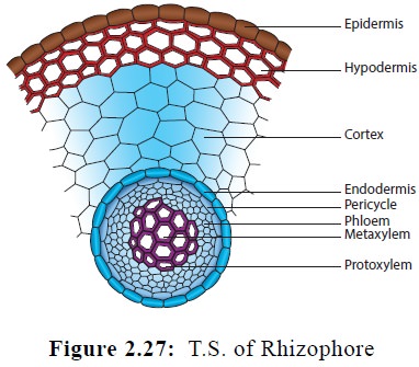
Rhizophore
The outermost layer of Rhizophore is the epidermis.
It is single layered and is covered with a thick cuticle. The cortex is differentiated
into outer scelrenchymatous and inner parenchymatous layers.
The innermost layer of cortex forms endodermis. The
stele is a protostele Figure 2.27. It is monarch and exarch but it is
centrifugal in S. kraussiana and
crescent shaped in S. atroviridis.
Stem
The anatomy of the stem reveals the presence of
epidermis, cortex and stelar region (Figure 2.28).
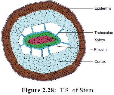
The epidermis is parenchymatous and is covered with
a thick cuticle. The cortex is parenchymatous with cells arranged without
intercellular spaces. A sclerenchymatous hypodermis is noticed in Selaginella lepidophylla. The presence
of radially elongated endodermal cell s called trabeculae is the characteristic feature of Selaginella. The casparian strips are found on the lateral walls.
The rapid stretching of the innermost cortical cells in comparison with stele
results in air spaces and stele appears to be suspended in air space with the
help of trabeculae. The stele is a protostele and exarch. A variation in number
of steles is found. It may be monostelic (S.
spinulosa); distelic (S. kraussiana
)or polystelic (S. laevigata). The
xylem is monarch(S. kraussiana) or
diarch (S. oregana) . Tracheids are
present but vessels are also noticed in S.
densa and S. rupestris.
Leaf
The leaf shows upper and lower epidermis. The
epidermal cells have chloroplast. Stomata occur on both surfaces. The mesophyll
is made up of loosely arranged thin walled cells with intercellular spaces.
There is a median vascular bundle surrounded by a bundle sheath. In vascular
bundle xylem is surrounded by phloem.
Reproduction
Selaginella
shows
both vegetative and asexual modes of
reproduction.
Vegetative reproduction
Selaginella
reproduces
vegetatively by fragmentation, bulbil
formation, tuber formation and resting buds.
Sexual reproduction
During
sexual reproduction spores are produced (Figure 2.29). Selaginella is
heterosporous and produces two types of spores namely microspores in microsporangium and megaspores in megasporangium.
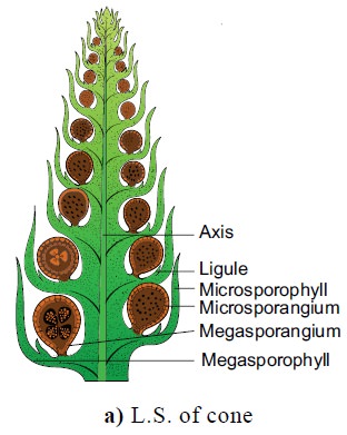
The sporangia are borne singly in the axils of microsporophyll and
megasporophyll respectively. The sporophylls are arranged spirally around a
central axis and aggregate to form strobili or cones. Variations in the
distribution of microsporangia and megasporangia among the species are seen. In
S. selaginoides and S. rupestris megasporangia are present
in the basal part of the cone. S. kraussiana possesses a single
megasporangium at the base of the strobilus. In S. inaequifolia one side of the strobilus bear only megasporangia
and other microsporangia. Separate strobili for microsporangia and
megasporangia are present in S. gracilis.
and S. atro-viridis.
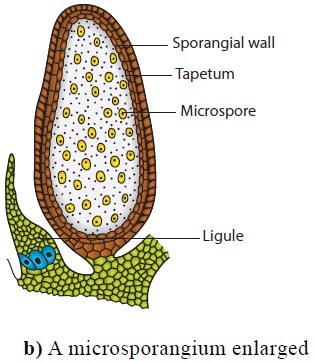
The development of sporangium is of eusporangiate
type. The sporangial initial divides periclinally to form outer jacket initials
and inner archesporial initials. The archesporial initials by repeated
anticlinal and periclinal divisions form sporogenous cells. Microspore mother
cells of microsporangiumundergoreductiondivision to produce halpoid
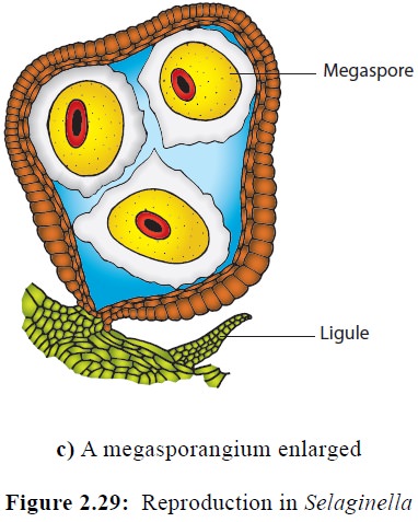
microspores. Similarly the megaspore mother cell undergoes reduction division
to produce 4 haploid megaspores. The microspore and megaspore represent the
male and female gametophyte and germinate inside the sporangium. The
microspores ![]()
![]()
![]() produce biflagellate
antherozoids. Archegonia develop in the megaspore. The antherozoids swim in
water and reach the archegonium. Fertilization brings the fusion of male and
female nucleus which result in the formation of a diploid zygote. The diploid
zygote represents the first cell of sporophyte.
produce biflagellate
antherozoids. Archegonia develop in the megaspore. The antherozoids swim in
water and reach the archegonium. Fertilization brings the fusion of male and
female nucleus which result in the formation of a diploid zygote. The diploid
zygote represents the first cell of sporophyte.
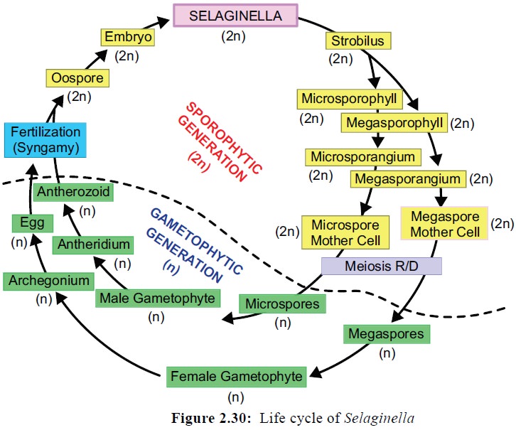
It undergoes several mitotic division to form
embryo. The embryo develops into a mature sporophytic plant.
In the life cycle of Selaginella alternation of
sporophytic and gametophytic generation is present (Figure 2.30).
Related Topics