Chapter: 11th Botany : Chapter 2 : Plant Kingdom
Cycas - Gymnosperms
Cycas
Class–Cycadopsida
Order – Cycadales
Family- Cycadaceae
Genus - Cycas
It is widely distributed in tropical and sub
tropical region of eastern hemisphere of the world. Cycas revoluta, Cycas
beddomei, Cycas circinalis, Cycas
rumphii are some of the common
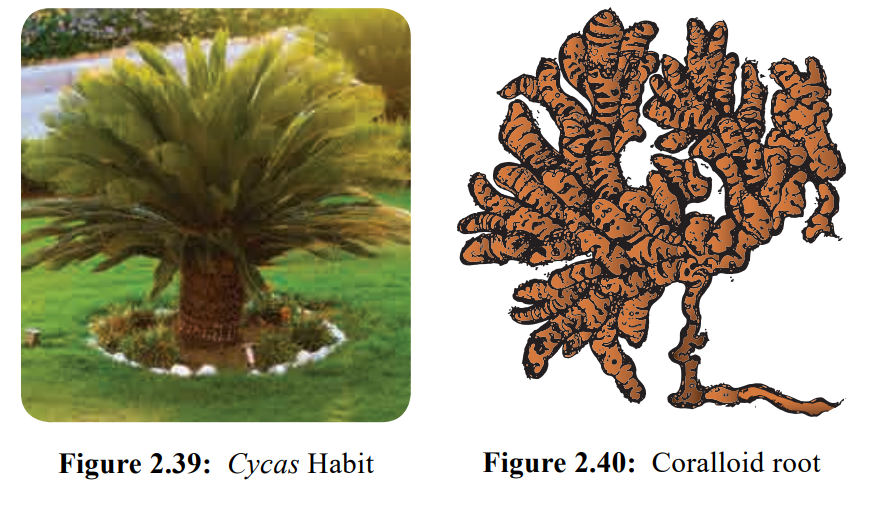
species. The plant body is sporophyte and resemble a small palm. The growth is
very slow. It is evergreen and xerophytic in nature.
Sporophyte:-
The sporophyte is differentiated into root, stem
and leaves. The stem is columnar bearing a crown of spirally arranged pinnately
compound leaves (Figure 2.39).
External features
Root
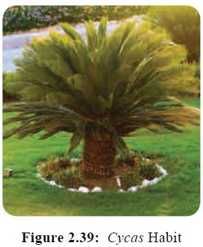
Two types of roots are found in Cycas. They are the tap root and
coralloid root.
The primary root persists and forms the tap root.
Some of the lateral roots give rise to branches which grow vertically upward
below the ground level. They branch repeatedly to form dichotomously branched
coral- like roots called coralloid roots. The cortical region of the coralloid
root contains the Blue green alga – Anabaena
sp. which helps in nitrogen fixation
(Figure 2.40).
Stem
The stem is columnar, unbranched and woody. It is
covered with persistent woody leaf bases. The stem also bears adventitious buds
at the base.
Leaves
Cycas has two
types of leaves
(i) Foliage
or assimilatory leaves
(ii) Scale
leaves
(i) Foliage or assimilatory leaves
Foliage leaves are large, pinnately compound and
form a crown at the top of the stem. Each leaf has 80-100 pairs of sessile leaflets.
The apex is acute or spiny. The leaflet has a single midvein. Lateral veins are
absent. Circinate vernation is present and young leaves are covered with ramenta.
(ii) Scale leaves
Scale leaves are brown, small, triangular and
persistent which are protective in function. They are covered with ramenta.
Internal structure
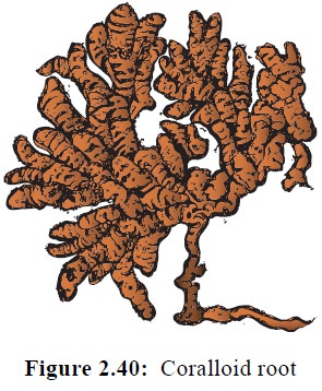
T.S. of Root
The internal organization of the primary root
reveals the following parts.
1. Epiblema, 2. Cortex 3. Vascular region (Figure
2.41). Epiblema is the outermost layer and is made up of single layered
parenchyma. It is followed by thin walled parenchymatous cortex. The cortex is
delimited by single layered endodermis. A multilayered parenchymatous pericycle
is present and it surrounds the vascular tissue. The xylem is diarch in young
root and tetrarch in older ones. Secondary growth is present. Coralloid root
also shows similar structure but the middle cortex is characterized by the
presence of Algal zone. Blue green alga called, Anabaena is found in
this zone. The xylem is triarch and
exarch.
T.S. of Stem
The cross section of young stem is irregular in
outline due to the presence of persistent leaf bases. It is differentiated into
epidermis, cortex and vascular cylinder. It resembles the structure of a dicot
stem (Figure 2.42).
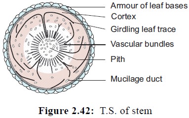
The epidermis is the outermost layer and is covered
with thick cuticle. It is discontinuous due to the presence of leaf bases. The
cortex constitutes the major part and is made up of thin walled parenchymatous
cells. The cells are filled with starch grains. Cortex also possesses several
mucilage ducts and tannin cells. In young stem the vascular bundles are
arranged in the form of a ring. A broad medullary ray is present. The vascular
bundles are conjoint, collateral, endarch and open. Xylem is made up of
tracheids and phloem consists of sieve tubes and phloem parenchyma. Companion
cells are absent. The cambium present in the vascular bundle is active for
short period. The secondary cambium is formed from the pericycle or cortex and
helps in secondary growth of the stem. The cortical region shows a large number
of leaf traces. The presence of direct leaf traces and girdling leaf trace is
the unique feature of Cycas stem.
Secondary growth results in polyxylic
condition. Phellogen and cork are formed and replace the epidermis.The wood
formed belongs to manoxylic type.
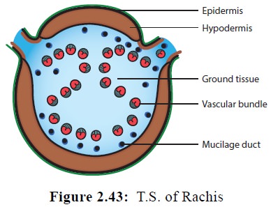
T.S. of Rachis
The outermost layer is epidermis and is covered by
thick cuticle. The hypodermis is made up of two layers of sclerenchyma on the
adaxial side and many layered on the abaxial side. The ground tissue is
parenchymatous. The peculiar feature of the rachis is the arrangement of
vascular bundle i.e., in an inverted Omega shape pattern (Figure 2.43). Each
vascular bundle is covered by a single layered sclerenchymatous bundle sheath.
Vascular bundles are collateral, endarch and open. A single layered endodermis
and few layered pericycle surrounds the bundle. A diploxylic condition is
present in the vascular bundles.( presence of both centripetal and centrifugal
xylem).
T.S. of Leaflet
The leaflet of Cycas in transverse section shows the presence of upper and lower epidermis. The epidermal cells are thick walled and are covered with thick cuticle. The lower epidermis is not continuous and is interrupted by sunken stomata. The hypodermis consists of sclerenchyma cells to prevent transpiration. The mesophyll is differentiated into palisade and spongy parenchyma.
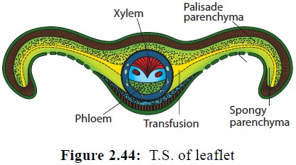
The cells of this layer are involved in
photosynthesis. The spongy parenchyma present in close proximity to the lower
epidermis bear large intercellular spaces which help in gaseous exchange.
![]()
![]()
![]()
Layers of colourless, elongated cells which run
parallel to the leaf surface from the midrib to the margin of the leaflet are
seen. These constitute the Transfusion
tissue that helps in the lateral
conduction of water. The vascular
bundle has xylem facing upper epidermis and phloem facing lower epidermis. The
protoxylem occupies the centre, hence the bundle is mesarch. The vascular
bundle has a sclerenchymatous bundle sheath (Figure 2.44).
Reproduction
Cycas reproduces
by both vegetative and sexual methods
Vegetative reproduction
It takes place by adventitious buds or bulbils.
They develop in the basal part of the stem. The bulbils on germination produce
new plants.
Sexual reproduction
Cycas is
dioecious i.e., male and female cones
are produced in separate plants. It is heterosporous and produces two types of
spores (Figure 2.45).
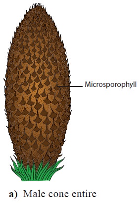
Male cone
The male cone or staminate cone are borne singly on
the terminal part of the stem. The growth of the stem is continued by the
formation of axillary buds at the base of the cone. The male cone is displaced
to one side showing sympodial growth in the stem. Male cones are stalked,
compact, oval or conical and woody in structure. It consists of several
microphylls which are arranged spirally around a central cone axis.
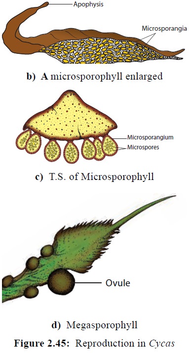
Microsporophylls
![]()
![]()
![]()
Microsporophylls are flat, leaf-like and woody
structures with narrow base and expanded upper portion. The upper expanded
portion becomes pointed and is called apophysis. The narrow base is attached to
the cone axis. Each microsporophyll contains thousands of microsporangia in
groups called sori on abaxial (lower) surface. Development of sporangium is of
Eusporangiate type. The spore mother cell undergoes meiosis to produce halpoid
microspores. Each Microsporangium bears large number of microspores or pollen
grains. Each sporangium is provided with a radial line of dehiscence, which
helps in the dispersal of spores. Each microspore (Pollen grain) is a rounded,
unicellular and uninucleate structure surrounded by outer thick exine and an
inner thin intine. The microspore represents the male gametophyte.
Megasporophylls
The megasporophylls of Cycas are not organised into cones. They occur in close spirals
around the tip of the stem of female plant. The megasporophylls are flat and
measuring 15 -30 cm in length. Each megasporophyll is differentiated into a
basal stalk and an upper leaf like portion. The ovules are attached to the
lateral side of the sporophyll. The ovules contain megaspore and it represent
the female gametophyte.
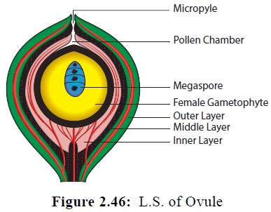
Structure of Ovule
Cycas produces
the largest ovule of the plant
kingdom. The ovules are orthotropous, unitegmic and possess a short stalk. The
single integument is very thick and covers the ovule leaving a small opening
called micropyle. The integument
consists of 3 layers, the outer and inner are fleshy (sarcotesta), the middle layer is stony called sclerotesta. The inner layer remains fused with the nucellus. The
nucellus grows out into a beak-like structure and the upper part dissolves and
forms a cavity-like structure called pollen
chamber. A single megaspore mother
cell undergoes meiosis to form four
haploid megaspores. The lowermost becomes functional and others get
degenerated. The nucellus gets reduced in the form of a thin papery layer in
mature seeds and encloses the female gametophyte An enlarged megaspore or the
embryo-sac is present within the nucellus. An archegonial chamber with 3-6
archegonia are present in the archegonial chamber below the pollen chamber
(Figure 2.46).
Pollination and Fertilization.
Pollination is carried out by wind and occurs at 3
celled stage(a prothallial cell, a large tube cell and a small generative
cell). Pollen grains gets lodged in the pollen chamber after pollination. The
generative cell divides into a stalk and a body cell. The body cell divides to
produce two large multiciliated antherozoids or sperms. During fertilization,
one of the male gamete or multiciliated antherozoid fuses with the egg of the
archegonium to form a diploid zygote (2n). The endosperm is haploid. The
interval between pollination and fertilization is 4- 6 months. The zygote
undergoes mitotic division and develops into embryo. The ovule is transformed
into seed. The seed has two unequal cotyledons. Germination is hypogeal. The
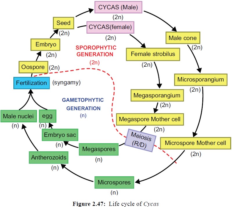
life cycle shows alternation of generations (Figure 2.47).
Related Topics