Chapter: Ophthalmology: The Eyelids
Eyelids: Basic Knowledge
Basic Knowledge
Protective function of the eyelids: The eyelids are folds of muscular softtissue that lie anterior
to the eyeball and protect it from injury. Their shape is such that the eyeball is completely covered when they are
closed. Strong mechanical, optical, and acoustic stimuli (such as a foreign
body, blinding light, or sudden loud noise) “automatically” elicit an eye closing reflex. The cornea is also
protected by an additional upward movement of the eyeball (Bell’s phenomenon). Regular
blinking (20 – 30 times a minute) helps to uni-formly distribute glandular
secretions and tears over the conjunctiva and cor-nea, keeping them from drying
out.
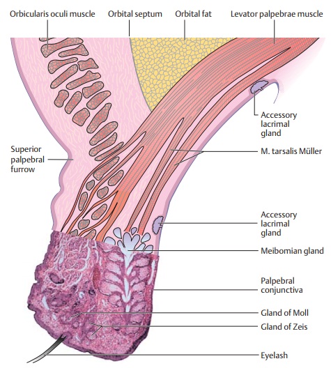
Structure of the eyelids: The eyelids consist of superficial and deep layers(Fig. 2.1).
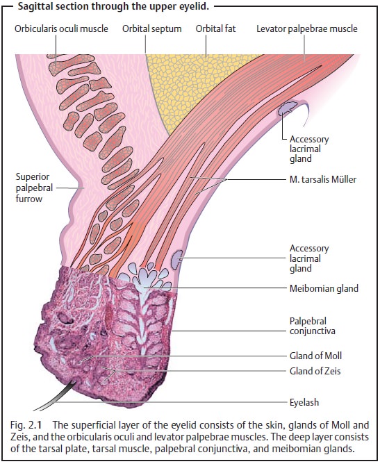
❖Superficial layer:
– Thin,
well vascularized layer of skin.
– Sweat
glands.
– Modified sweat gland and sebaceous glands (ciliary glands or glands ofMoll) and sebaceous glands (glands of Zeis) in the vicinity of the
eye-lashes.
– Striated muscle fibers of the orbicularis oculi muscle that actively closes the eye (supplied by the facial nerve).
❖ Deep layer:
– Thetarsal plate gives the eyelid firmness and shape.
– Smooth musculature of the levator palpebrae
that inserts into the tarsal plate (tarsal muscle). The tarsal muscle is supplied by the sympathetic nervous
system and regulates the width of the palpebral fissure. High sympathetic tone
contracts the tarsal muscle and widens the palpebral fissure; low sympathetic
tone relaxes the tarsal muscle and narrows the palpebral fissure.
– The palpebral conjunctiva is firmly attached to the tarsal plate. It forms an articular layer for the eyeball. Every time the eye blinks, it acts like a windshield wiper and uniformly distributes glandular secretions and tears over the conjunctiva and cornea.
– Sebaceous glands (tarsal or meibomian glands), tubular structures in the cartilage of the
eyelid, which lubricate the margin of the eyelid. Their function is to prevent
the escape of tear fluid past the margins of the eyelids. The fibers of
Riolan’s muscle at the inferior aspect of these sebaceous glands squeeze out
the ducts of the tarsal glands every time the eye blinks.
The eyelashes project from the anterior aspect of the
margin of the eyelid. On the upper eyelid, approximately 150 eyelashes are
arranged in three or four rows; on the lower eyelid there are about 75 in two
rows. Like the eyebrows, the eyelashes help prevent dust and sweat from entering the
eye. The orbital septum is located between the tarsal plate and the margin of
the orbit. It is a membranous sheet of connective tissue attached to the margin
of the orbit that retains the orbital fat.
Related Topics