Chapter: Ophthalmology: The Eyelids
Eyelids: Benign Tumors
Tumors
Benign Tumors
Ductal Cysts
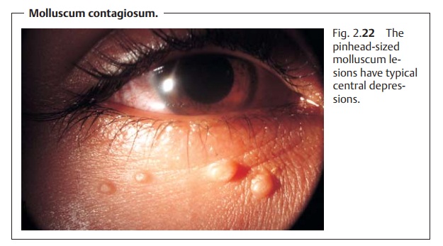
The round cysts of the glands or Moll are usually located in the angle of theeye. Their contents are clear and watery and can
be transilluminated. Gravitycan result in ectropion (Fig. 2.20). Therapy consists of marsupialization. The prognosis is good.
Xanthelasma
Definition
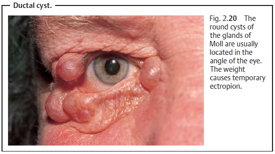
Local fat metabolism disorder that produces lipoprotein deposits. These are usually bilateral in the medial canthus.
Epidemiology:
Postmenopausal women are most frequently
affected. Ahigher incidence has also been observed in patients with diabetes,
increased levels of plasma lipoprotein, or bile duct disorders.
Symptoms:
The soft yellow white plaques are sharply demarcated. They areusually bilateral and distributed symmetrically (Fig. 2.21). Aside from the cos-metic flaw, the patients are asymptomatic.
Treatment and prognosis:
The plaques can only be removed surgically.
Theincidence of recurrence is high.
Molluscum Contagiosum
The noninflammatory
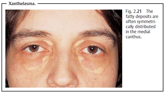
contagious infection is caused by DNA viruses. The disease usually affects children and teenagers and is
transmitted by direct contact. The pinhead-sized
lesions have typical central depressions and are scattered near the upper
and lower eyelids (Fig. 2.22). These lesions are re-moved with a curet. (In children this is
done under short-acting anesthesia.)

Cutaneous Horn
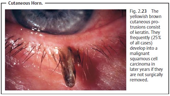
The yellowish brown cutaneous
protrusions consist of keratin
(Fig. 2.23). Older patients are more frequently affected. The cutaneous
horn should be surgically removed as 25% of keratosis cases can develop into malignantsquamous cell carcinomas years
later.
Keratoacanthoma
A rapidly growing tumor with a central
keratin mass that opens on the skinsurface, which can sometimes be expressed (Fig. 2.24). The tumor mayresolve spontaneously, forming a small sunken
scar.
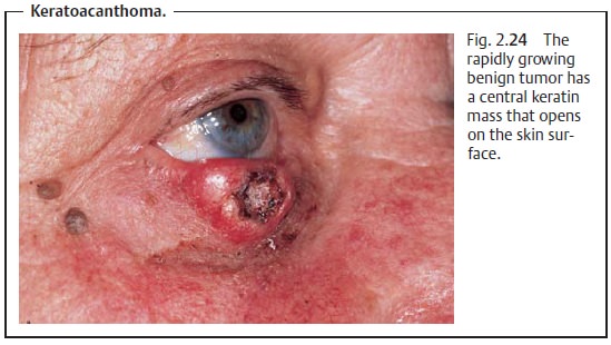
Differential diagnosis should exclude abasal cell
carcinoma(see that section);the margin of a keratoacanthoma is
characteristically avascular. Likewise, a squamous
cell carcinoma can only be excluded by a biopsy.
Hemangioma
Definition
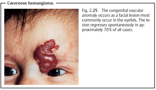
Congenital benign vascular anomaly resembling a neoplasm that is most frequently noticed in the skin and subcutaneous tissues.
Epidemiology:
Girls are most often affected (approximately
70% of all cases).
Facial lesions most commonly occur in the eyelids (Fig. 2.25).
Symptoms:
Hemangiomas include capillary or superficial,
cavernous, anddeep forms.
Diagnostic considerations:
Hemangiomas can be compressed, and the
skinwill then appear white.
Differential diagnosis:
Nevus flammeus:This is characterized by a sharplydemarcated bluish red mark
(“port-wine” stain) resulting from vascular expansion under the epidermis (not
a growth or tumor).
Treatment:
A watch-and-wait approach is justified in
light of the high rate ofspontaneous
remission (approximately 70%). Where there is increased risk of amblyopia due to the size of the lesion, cryotherapy,
intralesional steroidinjections, or radiation therapy can accelerate regression
of the hemangioma.
Prognosis:
Generally good.
Neurofibromatosis (Recklinghausen’s Disease)
Definition
A congenital developmental defect of the
neuroectoderm gives rise to neural tumors and pigment spots (café au lait spots).
Neurofibromatosis is regarded as a phacomatosis (a developmental disorder involving the simultaneous presence of changes in the skin, central nervous system, and ectodermal portions of the eye).
Symptoms and diagnostic considerations:
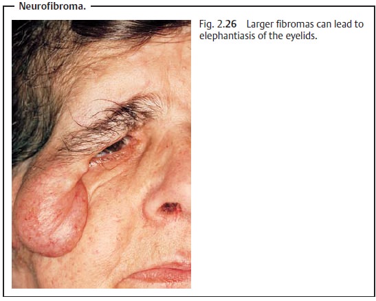
The numerous tumors are soft,broad-based, or
pediculate, and occur either in the skin or in subcutaneous tissue, usually in
the vicinity of the upper eyelid.
They can reach monstrous proportions and
present as elephantiasis of theeyelids (Fig.
2.26).
Treatment:
Smaller fibromas can be easily removed by
surgery. Largertumors always entail a risk of postoperative bleeding and
recurrence. On the whole, treatment is difficult.
Related Topics