Chapter: Ophthalmology: Retina
Retinal Dystrophies: Macular Dystrophies
Retinal Dystrophies
Macular Dystrophies
Definition
Macular dystrophies are disorders of the
macula that usually occur bilaterally and manifest themselves between the ages
of 10 and 30.
Stargardt’s Disease
Definition
This is a macular dystrophy that proceeds from
the retinal pigment epithelium.
Inheritance:
Autosomal recessive disorder.
Epidemiology:
Stargardt’s disease is rare.
Symptoms:
Progressive loss of visual acuity occurs between the ages of
10and 20 years.
Findings and diagnostic considerations:
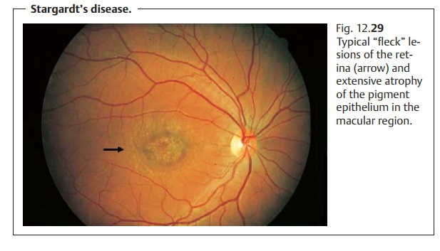
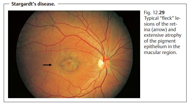
Initial findings are slight withwhite “fleck”
lesions in the macular region (Fig. 12.29),
which may occur in combination with lesions in the entire fundus (fundus flavimaculatus). The
electroretinogram and electro-oculogram will be normal or reduced. In the later
stage, the white lesions significantly increase in size and number. This will
not necessarily be reflected in the ERG or EOG.
Differential diagnosis:
Other disorders involving white “fleck” lesions suchas inherited
autosomal dominant drusen must by excluded by ophthalmos-copy. The diagnosis is
confirmed by fluorescein angiography. Blockage of the choroidal fluorescein is
a characteristic feature of Stargardt’s disease.
Treatment:
No treatment is available. Edge-filtered eyeglasses and magnify-ing near vision aids can help make better use of the patient’s remaining vision.
Prophylaxis:
No prophylaxis is possible. Examination of siblings and
geneticcounseling are indicated.
Clinical course and prognosis:
The disorder is chronically progressive.
Vision in the final stages is usually 0.1
(20/200) or less.
Best’s Vitelliform Dystrophy
Epidemiology:
The disorder is rare, with an incidence similar to
Stargardt’sdisease.
Inheritance:
The disorder is inherited as an autosomal dominant trait
withvariable penetrance and expressivity. The gene locus is on chromosome 11
(11q13).
Symptoms:
Clinical manifestation occurs between the ages of 5 and 15
years.Initially there is a subjectively slight decrease in visual acuity. In
the later stages of the disorder, vision is reduced to about 20/200.
Findings and diagnostic considerations:
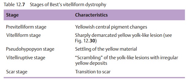
A typical feature of this form ofmacular dystrophy is that visual acuity is negligibly diminished at the onset ofthe disorder. However, the morphologic findings are remarkable. Ophthalmos-copy will reveal yellowish round vitelliform lesions in the macular region (Fig. 12.30) that look like the yolk of a fried egg. (The Latin word vitellus means egg yolk.) Usually these lesions are bilateral and symmetrical, although eccentric lesions may also occur. Table 12.7 lists the various manifestations.
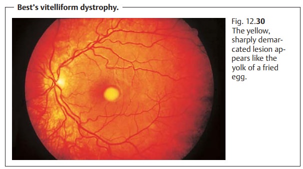
The macular change resembling an egg yolk gave rise to the name vitelliform dystrophy.
Differential diagnosis:
An unequivocal diagnosis can usually be made on thebasis of the
clinical picture alone. Sharply reduced or absent light response in the EOG and
ERG confirms the presence of Best’s vitelliform dystrophy.
Treatment:
The causes of the disorder cannot be treated.
Prophylaxis:
Examination of siblings and genetic counseling are indicated.
Clinical course and prognosis:
The prognosis is more favorable than for Star-gardt’s disease.
The disorder is chronically progressive. Visual acuity in the better eye
usually remains about 20/40. Secondary loss of visual acuity can result from
subretinal neovascularization.
Related Topics