Chapter: Ophthalmology: Retina
Central Serous Chorioretinopathy

Central Serous Chorioretinopathy
Definition
Serous detachment of the retina and/or retinal
pigment epithelium.
Etiology:
Serous detachment occurs through a defect in the outer blood –
ret-ina barrier (“tight junctions” in the retinal pigment epithelium). Local
factors that may be related to physical
or psychological stress are presumably involved.
Epidemiology:
The disorder primarily affects men in the third and fourthdecade
of life.
Symptoms:
Patients present with a loss of visual acuity, a relative
central sco-toma (dark spot), image distortion (metamorphopsia), or perception
of objects as larger or smaller than they are (macropsia or micropsia).
Diagnostic considerations:
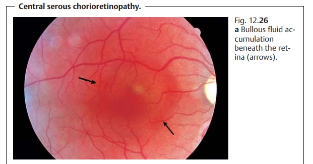
Ophthalmoscopywill reveal a serous retinaldetachment,
usually at the macula. In chronic cases, a fine brown and white pigment
epithelial scar will develop at the site of the fluid effusion. Swelling in the
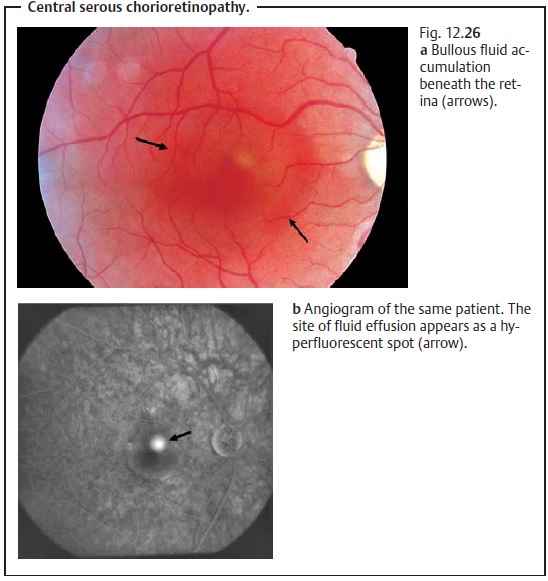
central retina shortens the visual axis and produces hyperopia. The site of
fluid effusion can be identified during the active phase with the aid of fluorescein angiography (Fig. 12.26a and b).

Treatment:
Usually no treatment is required for thefirst occurrenceof thedisorder. Retinal swelling resolves spontaneously within a few weeks. Recur-rences may be treated with laser therapy provided the site of fluid effusionlies outside the fovea centralis. Corticosteroid therapy is contraindicated as the therapy itself can lead to development of central serous chorioreti-nopathy in rare cases.
Clinical course and prognosis:
The prognosis is usually good. However,recurrences or chronic forms
can lead to a permanent loss of visual acuity.
Local stress-related factors and steroids can
lead to macular edema in predisposed patients.
Related Topics