Chapter: Ophthalmology: Retina
Degenerative Retinoschisis

Degenerative Retinoschisis
Definition
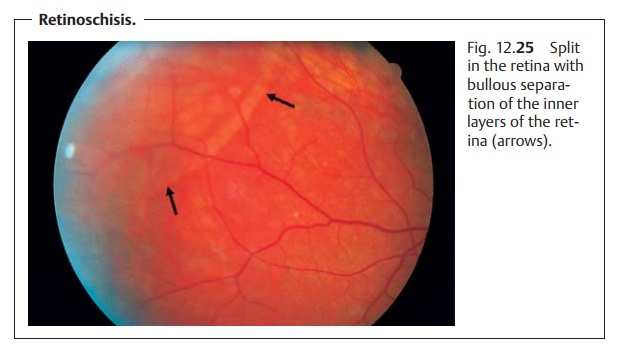
A frequently bilateral split in an inner and
outer layer of the retina. The split is usually at the level of the outer
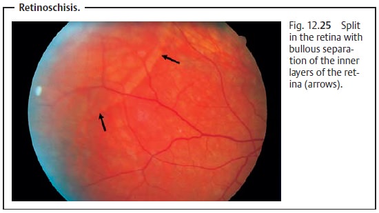
plexiform layer (Fig. 12.25).

Epidemiology:
About 25% of all people have retinoschisis. The
tendencyincreases with age.
Pathogenesis:
Idiopathic retinal splitting occurs, usually in the outer
plexi-form layer.
Symptoms:
Retinoschisis isprimarily
asymptomatic. The patient will usuallynotice a reduction of visual acuity
and see shadows only when the retinal split is severe and extends to the
posterior pole.
Diagnostic considerations:
Ophthalmoscopic examination will revealbullous separation of the split inner layer of the retina. The inner surface has the appearance of hammered metal. Rarely breaks will occur in the inner and outer retinal layers.
Differential diagnosis:
Rhegmatogenous retinal
detachmentshould be
ex-cluded. Ophthalmoscopy will reveal a continuous break in the retina in a
reti-nal detachment, and the retina will not appear as transparent as in
retino-schisis. However, retinal breaks can also occur in retinoschisis. In the
inner layer of the retina, these breaks will be very small and hardly
discernible. In the outer layer, they will be very large. Complete rhegmatogenous
retinal detachment can occur in retinoschisis only where there is a break in
both layers.
Treatment:
Usually no treatment is required. The rare cases in which
retinaldetachment occurs are treated surgically using the standard procedures
for retinal detachment.
Degenerative retinoschisis differs from
retinal detachment in that it usually requires no treatment.
Clinical course and prognosis:
The prognosis for degenerative retinoschisisis very good.
Progressive retinal splitting or retinal detachment with a sub-sequent
reduction in visual acuity is rare.
Related Topics