Chapter: Ophthalmology: Retina
Examination of the Fundus
Examination Methods
Visual Acuity
Examination of the Fundus
Direct ophthalmoscopy (Fig. 12.4a; see also Fig. 1.13):
A direct ophthalmo-scope
is positioned close to the patient’s eye. The examiner sees a 16-power
magnified image of the fundus.
Advantages.The high magnification permitsevaluation of small retinal find-ings such as diagnosing retinal microaneurysms. The dial of the ophthalmo-scope contains various different plus and minus lenses and can be adjusted as necessary. These lenses compensate for refractive errors in both the patient
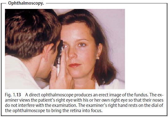
and the examiner. They may also be used to measure the prominence of retinalchanges, such as the prominence of the optic disk in papilledema or the prom-inence of a tumor. The base of the lesion is brought into focus first and then the peak of the lesion. A difference of 3 diopters from base to peak corre-sponds to a prominence of 1 mm. Direct ophthalmoscopy produces an erect image of the fundus, which is significantly easier to work with than an inverted image, and is therefore a suitable technique even for less experienced examiners.
Disadvantages.The image of the fundus is highly magnified but showsonly asmall portion of the fundus.
Rotating the ophthalmoscope can only partiallycompensate for this disadvantage.
Direct ophthalmoscopy also produces only a two-dimensional image.
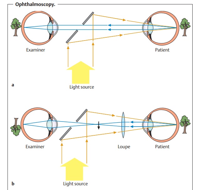
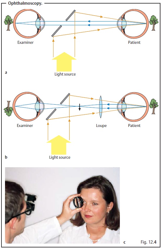
Indirect ophthalmoscopy (Figs. 12.4bandc):
A condensing lens (+ 14 to +
30diopters) is held approximately 13 cm from the patient’s eye. The fundus
appears in two to six-power magnification; the examiner sees a virtual inverted
image of the fundus at the focal point of the loupe. Light sources are
available for monocular or binocular examination.
Advantages.This technique provides a good stereoscopic, optimally
illumi-nated overview of the entire fundus in binocular systems.
Disadvantages.Magnification is significantly less than in direct
ophthalmos-copy. Indirect ophthalmoscopy requires practice and experience.
Contact lens examination:
The fundus may also be examined with a slitlamp when an additional magnifying lens such as a three-mirror lens (see Fig. 12.5) or a 78 to 90 diopter lens is used.
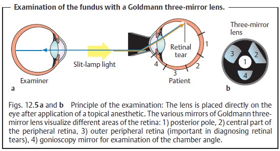
Advantages.This technique produces a highly magnified
three-dimensionalimage yet still provides the examiner with a good overview of
the entire fun-dus. The three-mirror lens also visualizes “blind areas” of the
eye such as the angle of the anterior chamber. Contact lens examination
combines the advan-tages of direct ophthalmoscopy and indirect ophthalmoscopy
and is there-fore the gold standard
for diagnosing retinal disorders.
Where significant opacification of the optic
media (as in a mature cata-ract) prevents direct visualization of the retina
with the techniques mentioned above, the examiner can evaluate the pattern of
the retinal vasculature. The sclera is directly illuminated in all four
quadrants by moving a light source back and forth directly over the sclera.
Patients with intact retinas will be able to perceive the shadow of their own
vasculature on the retina (entoptic phenomenon). They will see what looks like
“veins of a leaf in autumn”. Patients who are able to perceive this phenomenon
have potential retinal vision of at least 20/200.
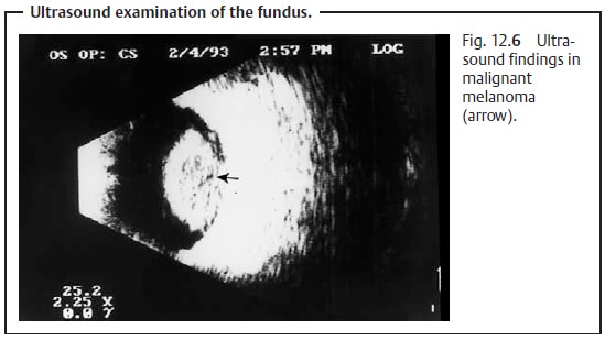
Ultrasonography:
Ultrasound studies are indicated where opacification ofthe optic
media such as cataract or vitreous hemorrhage prevent direct inspection of the
fundus or where retinal and choroidal findings are inconclu-sive. Intraocular tissues
vary in how they reflect ultrasonic waves. The retina is highly reflective,
whereas the vitreous body is normally nearly anechoic. Ultrasound studies can
therefore demonstrate retinal detachment and distin-guish it from a change in
the vitreous body. Optic disk drusen are also highly reflective. Ultrasound is
also helpful in diagnosing intraocular tumors with a prominence of at least 1.5
mm. The specific echogenicity of the tissue also helps to evaluate whether a
tumor is malignant, for example in distinguishing a choroidal nevus from a
malignant melanoma (Fig. 12.6).
Ultrasound studies can demonstrate retinal
detachment where the optic media of the eye are opacified (due to causes such
as cataract or vitreous hemorrhage). This is because the retina is highly
reflective in contrast to the vitreous body. Ultrasound can also be used to
confirm the presence of malignant choroidal processes.
Fundus photography:
Abnormal changes can be recorded with a single-lensreflex
camera. This permits precise documentation of follow-up findings. Photographs
obtained with a fundus camera in green light provide high-con-trast images of
abnormal changes to the innermost
layers of the retina such as changes in the layer of optic nerve fibers,
bleeding, or microaneurysms.
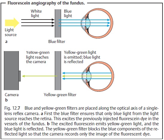
Fluorescence angiography (with fluorescein or indocyanine green):
Influorescein angiography, 10 ml of 5% fluorescein sodium are injected into one of the patient’s cubital veins. Blue and yellow-green filters are then placed along the optical axis of a single-lens reflex camera. The blue filter ensures that only blue light from the light source reaches the retina. The yellow-green filter blocks the blue components of the reflected light so that the camera records only the image of the fluorescent dye (Fig. 12.7).
Fluorescein angiography is used to diagnose
vascular retinal disorders such as proliferative diabetic retinopathy, venous
occlusion, age-related macular degeneration, and inflammatory retinal
processes. Where the blood-retina barrier formed by the zonulae occludentes is
disturbed, fluorescein will leak from the retinal vessels. Disorders of the
choroid such as choroiditis or tumors can also be diagnosed by this method; in
these cases indocyanine is better than fluorescein.
Related Topics