Chapter: Ophthalmology: Retina
Degenerative Myopia

Degenerative Myopia
Definition
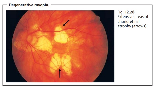
The fundus in degenerative myopia is characterized
by abnormal chorioretinal atrophy.
Epidemiology:
Chorioretinal atrophy due to myopia is rare.
Pathogenesis:
The atrophy usually occurs in the presence of severe
myopiaexceeding minus 6 diopters. The causes include stretching changes in the
ret-ina, choroid, and Bruch's membrane due to the elongated globe in axial
myopia.
Symptoms:
Loss of visual acuity occurs where there is macular involvement.
Findings and diagnostic considerations:
Typical signs include chorioretinalatrophy
around the optic disk and at the posterior pole and defects in Bruch’s membrane
known as lacquer cracks (Fig. 12.28).
These cracks can provide openings for vascular infiltration with resulting
subretinal neovasculariza-tion that can lead to retinal edema and bleeding
(Fuchs’ black spot). The final stage of the disorder is characterized by a
diskiform scar. The diagnosis is made by ophthalmoscopy. Fluorescein
angiography is indicated where sub-retinal neovascularization is suspected.

Differential diagnosis:
Choroidal scars and angioid streaks (breaks in Bruch’smembrane) in pseudoxanthoma elasticum must be excluded by ophthalmos-copy. The diagnosis is unequivocal where myopia is present.
Treatment:
The causes of the disorder cannot be treated. It is important
tocorrect myopia optimally with eyeglasses or contact lenses to avoid fostering
progression of the disorder. Subretinal neovascularization outside the fovea or
close to its border can be treated by laser photocoagulation.
Clinical course and prognosis:
Chronic progressive myopia will result inincreasing loss of visual acuity. The prognosis for subretinal neovasculariza-tion is poor. The incidence of retinal detachment is higher in myopic eyes.
Related Topics