Optical Instruments - Compound microscope | 12th Physics : UNIT 7 : Wave Optics
Chapter: 12th Physics : UNIT 7 : Wave Optics
Compound microscope
Compound microscope
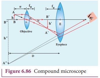
The diagram of a compound microscope is shown in Figure 6.86.
The lens near the object, called the objective, forms a real, inverted,
magnified image of the object. This serves as the object for the second lens
which is the eyepiece. Eyepiece serves as a simple microscope that produces finally
an enlarged and virtual image. The first inverted image formed by the objective
is to be adjusted close to, but within the focal plane of the eyepiece so that
the final image is formed nearly at infinity or at the near point. The final
image is inverted with respect to the original object. We can obtain the
magnification for a compound microscope.
Magnification of compound microscope
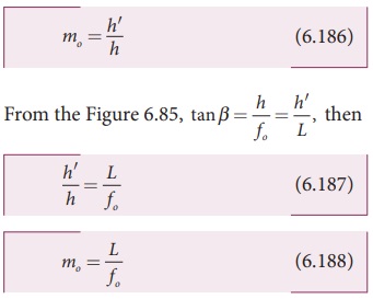
From the ray diagram, the linear magnification due to the
objective is,

From the Figure 6.85, tan ╬▓
= h/f0 = hŌĆ▓/L , then

Here, the distance L is between the first focal point of
the eyepiece to the second focal point of the objective. This is called the
tube length L of the microscope as fo and fe are comparatively smaller than L.
If the final image is formed at P (near point focussing), the
magnification me of the
eyepiece is,

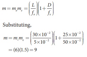
The total magnification m in near point focusing is,

If the final image is formed at
infinity (normal focusing), the magnification me of the eyepiece is,

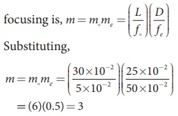
The total magnification m in normal focusing is,

EXAMPLE 6.42
A microscope has an objective and
eyepiece of focal lengths 5 cm and 50 cm respectively with tube length 30 cm.
Find the magnification of the microscope in the (i) near point and (ii) normal
focusing.
Solution
f0 = 5cm = 5├Ś10ŌłÆ2 m; fe
= 50cm = 50├Ś10ŌłÆ2 m;
L = 30cm = 30├Ś10ŌłÆ2 m;
D = 25cm = 25├Ś10ŌłÆ2 m
(i) The total magnification m in near point focusing is, m = mome

(ii) The total magnification m in normal focusing is, m = mome

Related Topics