Chapter: The Massage Connection ANATOMY AND PHYSIOLOGY : Introduction to Anatomy and Physiology
Cellular Level of Organization
CELLULAR LEVEL OF ORGANIZATION
So far, we have viewed the body at the chemical level and reduced it to a collection of chemicals. Such fragmentation is useful in understanding the physical properties of the body and how the chemical struc-tures contribute to the property. However, the body is much more than a mixture of chemical compounds. It is a dynamic, living being with its functional unit, the cell, behaving like a miniature human, respond-ing to internal and external stimuli.
The human body has two classes of cells—somaticcells and sex cells. The somatic cells include all cellsother than ova and sperm.
Typically, cells are surrounded by a medium known as extracellular fluid. Of the extracellular fluid, the fluid that actually surrounds the cells (i.e., fluid not inside blood vessels) is known as the interstitial fluid. The inside of the cell is separatedform the interstitial fluid by the cell membrane, which plays many important roles—it serves as a physical barrier between the inside of the cell and the extracellular fluid; it controls the entry of nu-trients and other substances; and it contains spe-cial receptors that respond to specific stimuli and alter the functioning of the inside of the cell. Con- nections between cell membranes of adjacent cells help stabilize the tissue.
Cytoplasm
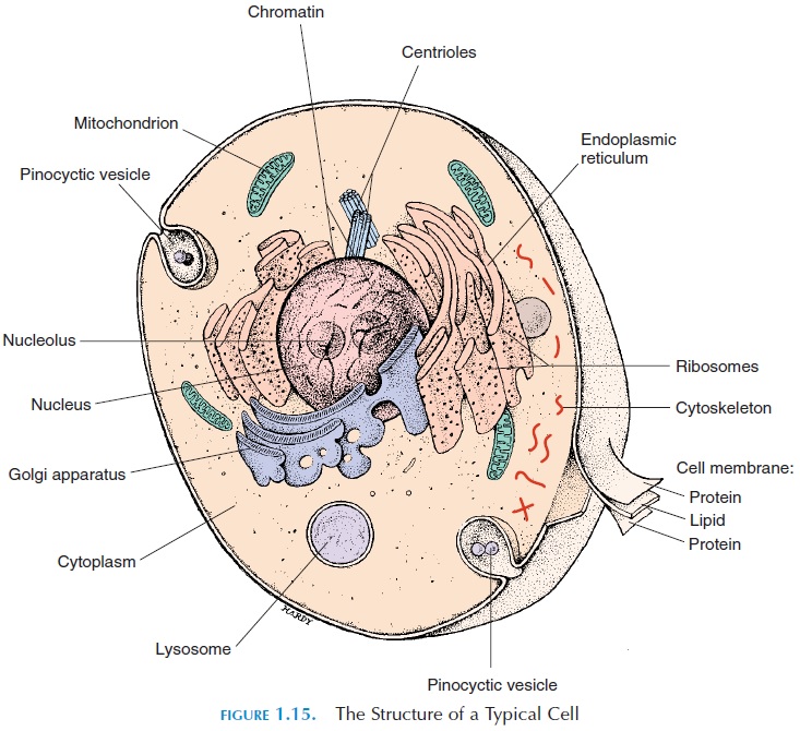
The material enclosed by the cell membrane is known as cytoplasm (see Figure 1.15). The cyto-plasm contains the nucleus and special structures, or organelles, floating in the fluid. The fluid inside thecell is known as the intracellular fluid, or cytosol.
Ions, soluble and insoluble proteins, and waste prod-ucts can be found in the cytosol. The major difference between the intracellular fluid and extracellular fluid is that the intracellular fluid has more potassium ions and proteins. In addition, it has stored nutrients in the form of glycogen and amino acids.
Organelles
The cell has many organelles. Some of the organelles are enclosed in a lipid membrane, separating them from the cytosol; others are in direct contact with the cytosol (Figure 1.15). The membranous organelles in-clude the mitochondria, endoplasmic reticulum, the Golgi apparatus, lysosomes, and peroxisomes.
The nonmembranous structures include the cy-toskeleton, the microvilli, centrioles, cilia, fla-gella, and ribosomes.
Mitochondria
The mitochondria are double-membrane structures, which may be long and slender or short and fat. The number of mitochondrion vary from cell to cell, from red blood cells, having no mitochondria, to liver cells, which are packed with them. The presence of mito-chondria indicates the demand for energy by specific cells.
The inner membrane of the mitochondria is thrown into folds to increase the surface area. The in-side of the mitochondria contains the enzymes re-quired for breaking down nutrients to liberate energy for cellular function; 95% of the ATP requirements are provided by mitochondrial activity.
Endoplasmic Reticulum
The endoplasmic reticulum, as the term suggests, is a network of intracellular membranes (which are in the form of tubes and sacs) that is connected to the nuclear membrane. It contains enzymes and partici-pates in protein and lipid synthesis. Some endoplas-mic reticulum appear smooth—smooth endoplasmic reticulum—while others appear rough as a result of the presence of ribosomes (discussed later).
The function of the endoplasmic reticulum varies from cell to cell. The rough endoplasmic reticulum helps manufacture, process, and sort proteins. Smooth endoplasmic reticulum helps manufacture fats and steroids. In muscle cells, it stores the cal-cium required for muscle contraction. In liver cells, it contains enzymes that help detoxify harmful agents such as drugs and alcohol.
Golgi Apparatus (Golgi Complex)
The Golgi apparatus appears as flattened membrane disks known as saccules. If considered similar to a factory, endoplasmic reticulum serves as the packag-ing center of the cell. Chemicals manufactured by the endoplasmic reticulum enter the Golgi complex where they are processed, sorted, and packaged in secretory vesicles ready for dispatch to the outside of the cell, or for storage inside the cell as storage vesi-cles. Secretions, such as hormones and enzymes, are packaged by this structure.
Lysosomes
Lysosomes are vesicles filled with digestive enzymes. The lysosomes are manufactured in the Golgi appa-ratus. The lysosomes enzymes are activated when they fuse with damaged organelles or other vesicles containing contents to be destroyed. On fusion, the enzymes become activated and digest the contents. Substances that can be recycled diffuse back into the cytosol. Unwanted contents are expelled into the ex-tracellular fluid by exocytosis. In sperm cells, lysoso-mal enzymes are secreted outside and help the sperm penetrate the ovum.
Peroxisomes
Peroxisomes are similar to lysosomes, except they help detoxify substances, such as alcohol and hydro-gen peroxide, that are produced by the cell. In this way, peroxisomes protect the cell from the harmful effects of toxic substances.
Cytoskeleton
The cytoskeleton is actually a framework of proteins located inside the cell that gives the cell its flexibility and strength. The cytoskeleton is in the form of fila-ments(threadlike structures) and tubules (micro-tubules). The filaments help anchor organelles inside the cell, as well as anchor the cells to surrounding ar-eas. Tubules help maintain the shape of the cell and help transport substances within the cell.
Microvilli
Microvilli are small fingerlike projections of the cell membrane that increase the surface area. They are found in those cells involved in absorbing substances from the extracellular fluid. Unlike the processes oc-curred by the cell membrane in endocytosis, the mi-crovilli are more stable and are anchored to the cy-toskeleton of the cell. The microvilli present on the surface of intestinal cells increase the surface area for absorption by 20%.
The Centrosome
The centrosome is a structure located close to the nu-cleus. It consists of the pericentriolar area, which is composed of protein fibers and centrioles. The cen-trioles are two, short, cylindrical structure composed of microtubules. They are only found in those cells capable of dividing. Muscle cell, neurons, mature red blood cells, and cardiac muscle cells—all cells not ca-pable of multiplying—lack centrioles. The centriole is important at the time of cell division to separate DNA material.
Cilia
Cilia are projections from the cell membrane found in certain cells, such as those in the respiratory tract. Cilia have nine pairs of microtubules, surrounding a central pair They move rhythmically in one direction and move mucus and other secretions over the cell surface.
Flagella
Flagella (singular, flagellum) can be considered longer cilia. Rather than moving the fluid over the cell surface like the cilia, flagella help move the cell in the surrounding fluid. A good example of a cell with flagellum is the sperm cell of the testis.
Ribosomes
Ribosomes are tiny organelles that manufacture pro-teins. They may be fixed to the endoplasmic reticu-lum (rough endoplasmic reticulum) or float freely in the cytosol.
The Nucleus
The nucleus of the cell is a denser area found in most cells (mature red blood cells do not contain a nu-cleus). Some cells, such as skeletal muscle cells, may have more than one nucleus. A membrane known as the nuclear membrane, or nuclear envelope, sur-rounds the nucleus, resembling the plasma mem-brane. Channels in the nuclear membrane control the movement of substances in and out of the nucleus. The nucleus contains a denser structure called the nucleolus, in which ribosomes (containing RNA) areassembled.
The nucleus contains all the information required for the cell to function and controls all cellular oper-ations. The nucleus has the information needed for the manufacture of more than 100,000 proteins. It also controls which proteins will be synthesized and in what amounts in a given time.
The information required by the cell is stored in DNA strands. The DNA strands are found in thread-like structures known as chromosomes. Each hu-man cell has 23 pairs of chromosomes.
DNA is actually a double-helix strand, with the two strands held together by hydrogen bonds (see Figure 1.16). The genetic code in the DNA is in the se-quence of nitrogenous bases. The nitrogenous bases adenine, thymine, cytosine, and guanine are ar-ranged in different ways to form the genetic code. Three of the bases, arranged in a specific way, code for a specific amino acid. In this way, the DNA has
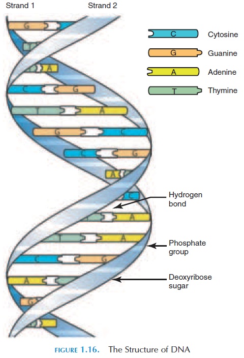
codes that give the sequence of arrangement of amino acids needed to form a specific protein. The lineup of bases that code for a specific protein is known as a gene, and a gene exists for every type of protein manufactured in the body.
When the gene is activated, it begins to manufac-ture proteins with the help of the ribosomes and RNA. The RNA carries the template of the genetic code to the cytoplasm and assures the amino acids are in the right sequence to form the protein.
Protein Synthesis
It should be noted that the proteins determine the characteristics of the cells, tissues, and the organism itself. Therefore, a large part of cellular activity is synthesizing different proteins; the instructions for the sequence of amino acids in the proteins are car-ried by the DNA.
The genetic code for a specific protein, present in the DNA, is used as a template to copy the sequence of amino acids for that protein. The copy is in the form of RNA. This process is called transcription. The RNA moves out of the nucleus into the cytoplasm. In the cy-toplasm, with the help of ribosomes and using the template on the RNA, amino acids are lined and bonded in the right sequence to form the specific pro-tein needed. This process is known as translation.
Cell Membrane (Plasma Membrane)
The cell membrane (see Figures 1.17and 1.18) is a thin, delicate layer that is made up of lipids, carbo-hydrates, and proteins. It is referred to as a phos-pholipid bilayerbecause it is made up of two layersof phospholipids. The phospholipids are lined in such a way that the end of the molecules containing the phosphate group that have an affinity for water— hydrophilic end—faces the outside of the cell mem-brane. The hydrophobic ends that contain the fatty acids face each other in the middle of the cell mem-brane. This arrangement of the cell membrane pre-vents water and water-soluble substances from cross-ing the lipid portion of the cell membrane. This arrangement is used because the composition of the cytoplasm of the cell is different from that of the fluid around it. These differences have to be maintained if the cell is to survive.
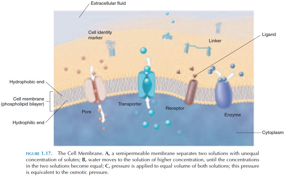
The phospholipid bilayer is interrupted in certain areas by proteins that go completely through the wall or are integrated into the wall with part of the protein molecule projecting into or out of the cell. These are known as membrane proteins. Because of the pres-ence of a large number of different proteins (mosaic) in the “sea” of phospholipids, the structure of the cell membrane is referred to as the fluid mosaic model.
The membrane proteins that go through and through are referred to as integral proteins. The others are referred to as peripheral proteins. The membrane proteins have many functions. Some pro-teins serve as anchors or linkers and connect the cell membrane to surrounding structures to stabilize the cell. Others serve as recognition proteins, or identi-fiers, or cell identity markers. These are usually gly-coproteins that project out of the membrane and help the immune cells identify the cell as self or nonself. Some of the peripheral proteins are enzymes and fa-cilitate chemical reactions inside or outside the cell, depending on their position; others are receptors.
Receptor proteins are specific and have an affinity for specific hormones and other substances. The spe-cific extracellular molecules that stimulate the recep-tors are referred to as ligands. Each cell may have re-ceptors for more than one ligand, and the receptors vary from cell to cell. In this way, hormones, which are carried throughout the body by the blood, affect only cells that have receptors for the specific hor-mone.
Certain proteins located in the cell membrane may serve as carriers or transporters. If a specific solute becomes attached to the carrier, the protein carrier changes shape and transports the solute across the cell membrane. This may occur with or without the use of active energy. Certain integral proteins work as channels or gates; forming small paths across thecell membrane and allowing water and specific ions to pass through. The channels may be opened by changes in potential or by binding of ligands.
The carbohydrates in the membrane, although only contributing about 3% of the weight of the cell membrane, project outward and help form a layer that protects the cell membrane.
Membrane Transport
The cell membrane determines which substances en-ter or leave the cell and is said to be impermeable if it does not allow any substance to pass through. A cell membrane can also be selectively permeable. It may be impermeable to one substance and freely al-low another to pass through. A typical cell membrane is selectively permeable. Because of its selective per-meability, the cell can maintain a different concen-tration of substances inside the cell than outside the cell. For example, there are more sodium ions out-side the cell compared with inside. This difference in chemical concentration is known as the chemicalgradient. If the inside and outside electrical chargesare compared, the inside of the cell is more negative than the outside. This is known as the electrical gra-dient. Many factors determine whether a substancecan pass through and the direction of movement.
Factors Affecting Transport
To some extent, the size of the substance plays a part, with the membrane being less permeable to those substances that are larger. The electrical charge of the substance has an effect on whether it is trans-ported. At rest, the inside of the cell is more negative than the outside. Substances that carry negative charges, therefore, find it more difficult to pass. The molecular shape of the substance also has an effect. Substances that are lipid-soluble pass through the membrane easily because the membrane is made up of phospholipids. The direction of movement is de-termined by the electrical and chemical gradients (electrochemical gradient). Transport may be af-fected by a combination of one or more factors.
The transport across the membrane may occur with or without the use of energy. Transport without use of energy is referred to as passive transport. For the transport of some substances, energy in the form of ATP must be used. This is known as active trans-port. In both of these transport types, transportersmay or may not be involved—known as mediated or unmediated transport, respectively. There are manymechanisms by which passive transport occurs.
Passive Transport
Five mechanisms are used for passive transport— diffusion, osmosis, filtration, carrier-mediated transport, and vesicular transport.
Diffusion
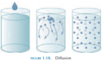
Diffusion (see Figure 1.18) is the movement of ions and molecules from an area of higher concentration to one of lower concentration. This difference in con-centration is known as the concentration gradient. Substances that are lipid-soluble diffuse directly through the phospholipid bilayer. Other substances, such as ions, diffuse through specific channels, if the channels are open. This passive process—diffusion— is important in the body. When the blood reaches the tissue, nutrients move from inside the blood vessels into the interstitial fluid by diffusion. The opposite also occurs by diffusion. Waste products from the cell move along the concentration gradient into the blood. Similarly, carbon dioxide and oxygen between the air and blood move by diffusion.
The rate of diffusion depends on the distance that separates the two solutions. To increase efficiency in the body, the distance of the cells from the blood is only about 125 micrometers ( m). The difference be-tween the concentrations of the two solutions also plays an important part. Oxygen in the body moves more rapidly into the tissue when the tissue has been active and the concentration of oxygen is much lower than in the blood.
Molecule size affects diffusion. Smaller particles tend to move at a faster pace than larger particles. Other than distance and concentration gradient and size, the electrical charges on the two substances af-fect diffusion, as the interior of the cell is negative. Even if a concentration gradient exists, a negatively charged substance finds it more difficult to enter the negatively charged cell. Temperature is another fac-tor that affects diffusion. Higher temperatures in-crease the diffusion rate.
Substances that are lipid-soluble, such as alcohol, fatty acids, and steroids, enter the cell easily through the lipid cell membrane if there is a concentration gradient. Substances that are water-soluble, however, must rely on the presence of channels to pass through, even if a concentration gradient exists. The surface area available for diffusion also determines the rate of movement. Because channels occupy only a small percentage of the cell membrane, diffusion through channels is comparatively slower than direct diffusion across the phospholipid bilayer.
Osmosis
Osmosis (see Figure 1.19) is the net diffusion of water from a region of lower concentration of solute (parti-cles) to a region of higher concentration of solute across a semipermeable membrane. Conversely, the movement of water from a region of higher concentra-tion (of water) to a region of lower concentration.
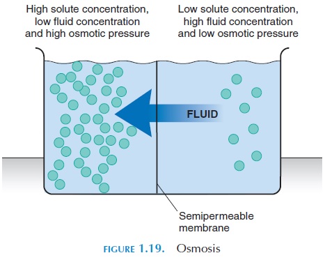
Three important characteristics of osmosis are:
1. Osmosis is the diffusion of water molecules across a membrane.
2. Osmosis occurs across a selectively permeable membrane that allows water to freely move through it; not the solutes.
3. The movement of water is toward the solution with the higher concentration of solutes.
In the body, the fluid inside the cell (intracellular fluid) and the fluid outside the cell (extracellular fluid) have dissolved substances. Each of these sub-stances tend to diffuse as if they were the only sub-stance in the solution. For example, if sodium and chloride are present, they each move along their own concentration gradient. The changes in the concen-tration gradient of chloride do not affect the move-ment of sodium.
In general, the total concentration remains the same on both sides. If the concentration of ions and molecules vary between the inside and outside of the cell, water is drawn by osmosis to the side that has more ions and molecules and less water.
Red blood cells can be used to illustrate osmosis. When placed in a glass of water, water rushes (by os-mosis) into the red blood cells because they have more particles inside. The cells swell and immedi-ately rupture. If the cells are placed in a glass of wa-ter into which two spoonsful of table salt was mixed, water from the cell moves out and the cells shrink. Osmotic pressure of a solution is an indication ofthe force of water movement into that solution as a result of its solute concentration.
Filtration
In filtration, water is forced across a semipermeable membrane as a result of hydrostatic pressure. For example, it is equivalent to the pressure that pushes water out of a nick in a garden hose through which water is flowing. By filtration, fluid moves out of cap-illaries. Similarly, fluid filtered from the blood into the renal tubules of the kidney finally form urine. The movement of larger particles, along with water, by fil-tration depends on the size of the pores present in the membrane.
Carrier-Mediated Transport
In this method of transport, integral proteins bind to specific ions, or other substances, and carry them across the cell membrane into the cell. Each carrier on the cell membrane is specific (i.e., it binds to only one specific substance). The amount of substance carried into the cell depends on the number of carri-ers present for that substance. Some carriers can carry two different substances. Both substances may be carried in the same direction or one substance may be carried out of the cell while the other is si-multaneously brought into the cell.
Substances, such as glucose and amino acids, are transported by carriers because they are insoluble in lipids and are too large to be transported through channels. Carriers specific for these substances bind to them and move them into the cell along the con-centration gradient. Here, no energy is used. It should be noted that it is a diffusion process, except that it is facilitated by carriers. This type of transport is referred to as facilitated diffusion. The rate at which they move into the cell depends on the number of carriers present on the cell membrane.
A unique property of carriers is that hormones can regulate them. Certain carrier activity is facilitated by the binding of hormones. In this way, hormones reg-ulate the movement of specific substances into the cell. For example, the hormone insulin facilitates the movement of glucose into the cell.
Vesicular Transport
With vesicular transport, vesicles or small mem-brane-lined sacs are used to bring substances into or out of the cell. The process of bringing substances in by forming vesicles is known as endocytosis. Trans-port of substances out of the cell in this manner is re-ferred to as exocytosis.
Endocytosis
Substances outside the cell that are too large to enter via channels are “engulfed” by a depression in the cell membrane. The cell membrane folds to form two processes, similar to two arms in an embrace, and the processes fuse with each other to form a vesicle inside the cytoplasm (see Figure 1.20A). In some types of endocytosis, the substance initially binds to receptor proteins before a vesicle is formed.
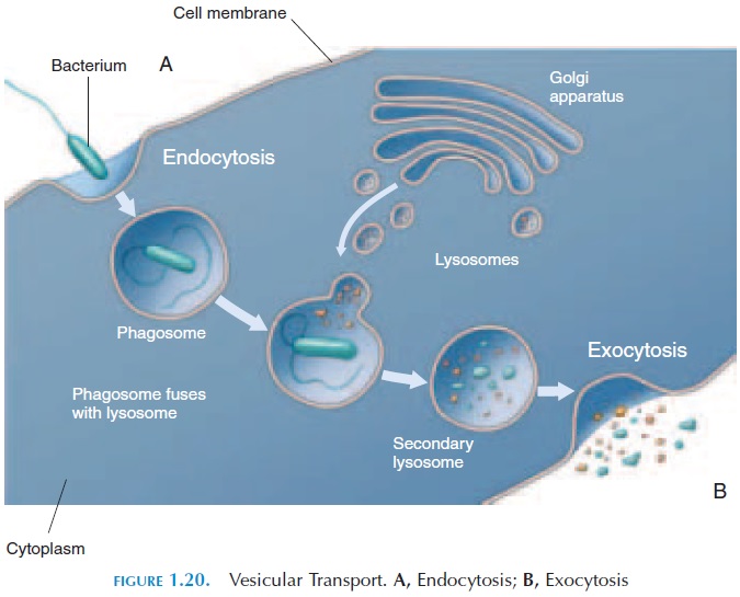
After endocytosis, at times, the contents are di-gested by enzymes (stored in vesicles) present in the cytoplasm. This process is known as phagocytosis (cell eating). Most defense cells kill microorganisms by phagocytosis.
Exocytosis
Exocytosis is the opposite of endocytosis (see Fig-ure 1.20B). Here, vesicles floating in the cytoplasm fuse with the cell membrane and extrude their con-tents into the extracellular fluid. Mucus secretion, se-cretory products of certain glands, and nerve endings extrude the contents of vesicles in this way.
Active Transport
Active transport (Figure 1.19) is the transport of sub-stances into or out of the cell using energy. Energy is needed for this kind of transport because it occurs against the concentration gradient, unlike diffusion. The carriers involved in this transport are referred to as ion pumps. All cells have specific ion pumps that transport sodium, potassium, calcium, and magne-sium. Ion pumps are specific (i.e., a pump is specific for one ion). There are certain pumps that transport one ion inside as another is sent outside. These spe-cial carrier proteins are known as exchange pumps. The most common exchange pump is the sodium–potassium exchange pump, or sodium–potassiumATPase.
Normally, the extracellular fluid has more sodium than the inside of the cell; potassium is the opposite. Sodium tends to diffuse in slowly along its concen-tration gradient, while potassium moves out. To maintain homeostasis, the sodium–potassium pump uses energy to pump out sodium and pump in potas-sium. This pump uses energy by consuming about 40% of the ATP produced in a resting cell.
Transmembrane Potential
All cells have more negative charges inside as com-pared with the outside. This difference in charges is maintained by the presence of a cell membrane that is selectively permeable and ionic pumps that move substances by active transport. This difference in electrical charge is known as the transmembranepotential. Transmembrane potential is measured inmillivolts (mV). The membrane potential of a neuron, for example, is 70 mV. The maintenance of trans-membrane potential is important, as it is required for many functions, such as transmission of nerve im-pulses, muscle contraction, and gland secretion.
The Cell Life Cycle
From fertilization to physical maturity, the cells un-dergo many divisions. When a single cell divides, it forms two daughter cells that are identical to the original cell. A cell may live from a few days to many years, depending on the cell type. Most cells have a gene, which is triggered to self-destruct at a specific time.
Cells divide in two ways: mitosis and meiosis. Mi-tosis is common and is the process of division seen in somatic cells, involving the separation of the dupli-cated chromosome into two identical nuclei. The cy-toplasm and the nucleus then separate into two new cells.
Meiosis can be seen in the testis and ovary during the formation of sperm and ova, in which the daugh-ter cells end up with half the number of chromo-somes found in somatic cells. When the ovum and sperm fuse during fertilization, the fused cell then has the right number of chromosomes.
When cells are not dividing, they continue to func-tion fully. This phase is known as the interphase. Cells that do not multiply after birth, such as neu-rons, are said to be in the interphase.
Cell division is regulated by peptides known as growth factors, which are present in the extracellularfluid. Growth factors bind to receptors in the cell membrane and trigger cell division. Growth hormone, nerve growth factor, epidermal growth factor, and ery-thropoietin are a few of the growth factors identified.
Specific genes, known as repressor genes, op-pose,cell division. When the rate of growth exceeds that of inhibition, the tissue enlarges. If uncontrolled cell growth occurs, a tumor or neoplasm results.
Related Topics