Chapter: Microbiology
Amoebiasis
Introduction
• Amoebiasis is an infection due to E.histolytica
• It occurs only among human and selected primates
• Commonly E.histolytica produce intestinal and extra intesti-nal infections
• Amoebiasis is transmitted by oral ingestion of materials con-taining cyst of E.histolytica
Morphology of the organism
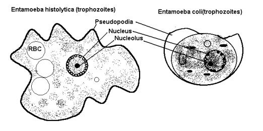
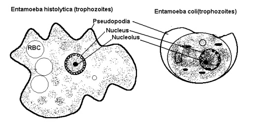
Trophozoites (Figure 22.1)
Entamoeba histolytica and entamoeba coli have both trophozoi-tes and cyst stages. The cytoplasms of E.histolytica is glossy, contains red cells and spherical vacuoles. The nucleus has small central karyo-some and fine regular chromatin granules lining the periphery of nuclear membrane. Entamoeba coli has granular cytoplasm which contains bacteria and other inclusions and ellipsoid vacuoles. Nucleus has ec-centric nucleolus and coarse beaded chromatin at the periphery.
Cysts
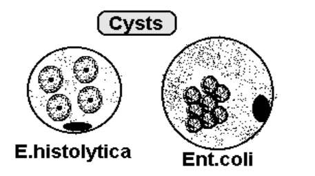
Cysts are round, nuclear morphology is similar to trophozoites. One to four nuclei are seen in E.histolytica and eight nuclei are present in Entamoeba coli cysts.
Clinical manifestations of the disease
• Both intestinal and extra intestinal amoebiasis have an incuba-tion period of more than one week (several weeks)
TYPES OF CLINICAL MANIFESTATONS
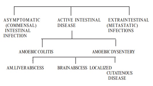
Asymptomatic Intestinal Infections
• Persons show no symptoms
• They pass cysts in stool
Active Intestinal Disease
• Minority of persons with intestinal infection develop diarrhoeal disease
• They pass red cells and pus in stool
• The symptoms and signs are:
- Less severe form
Fever
Abdominal pain
Tenesmus
- More sever form
Symptoms similar to ulcerative colitis
Peritonitis
Secondary intestinal perforations
Toxic mega colon
Pathology
· Parasites produce flask- shaped ulcers in which the base is wider than neck at the epithelial surface
· Organisms are present on the edge of the ulcer
Laboratory Diagnosis

Direct demonstration:
a. Wet mount:
1. Saline – Trophozoites,Cysts
2. Iodine – Cysts
3. LCB - Cysts
b. Stains : Iodine staining
: Iron haematoxylin stain
: Trichrome stain
: Immuno fluorescence staining
c. Culture: non-Axenic andAxenic culture methods are used
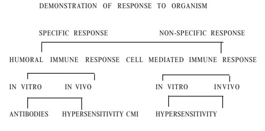
Indirect Methods
Epidemiology, Prevention and control
Cysts are ingested through contaminated food and water Flies transfer the cysts from infected stools to food
Control measures consist of improving environmental and food sanitation
Carriers must be barred from food handling
Metronidazole is the drug used for symptomatic amoebiasis
Related Topics