Chapter: Microbiology
The compound light microscope

THE COMPOUND LIGHT MICROSCOPE
Anton van Leeuwenhoek of Delft, Holland, constructed simple microscopes composed of double convex glass lenses held between two silver plates. His microscopes could magnify around 50 to 300 times. Microbiologists currently use a variety of light microscopes.
Modern microscopes are all compound microscopes. The light microscopy refers to the use of any kind of microscope that uses visible light to make the specimens observable. The most commonly used light microscopes are:
l Bright field microscopes
l Dark-field microscopes
l Phase contrast microscopes
l Fluorescence microscopes
The parts of a modern microscope and its light path are shown in figure. 2.1.
Each type of microscope is adapted for certain type of ob-servations. The standard ordinary light microscope is called a bright-field microscope, because it forms a dark image against a brighter background. A compound microscope with a single eye piece (ocular) is called monocular and with two eye pieces is called binocular.
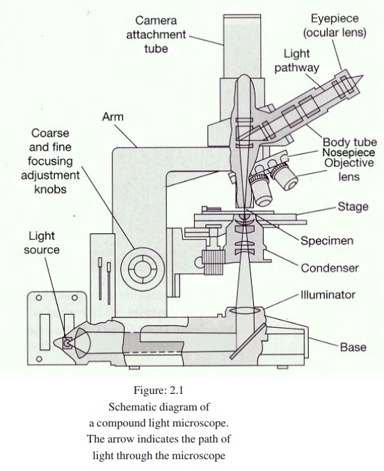
l A mirror or an electric illuminator is a light source which is located in the base of the microscope.
l There are two focusing knobs, the fine and the coarse adjust-ment knobs which are located on the arm. These are used to move either the stage or the nosepiece to focus the image.
l The mechanical stage is positioned about halfway up the arm, which allows precise contact of moving the slide.
l The sub stage condenser is mounted within or beneath the stage and focuses a cone of light on the slide. In simpler microscopes, its position is fixed whereas in advanced microscopes it can be adjusted vertically.
The upper part of the microscope arm holds the body assem-bly. The nose piece and one or more eyepieces or oculars are at-tached to it. The body assembly contains a series of mirrors and prisms so that the barrel holding the eyepiece may be tilted for viewing. Three or five objectives with different magnification power are fixed to the nose piece and can be rotated to the posi-tion beneath the body assembly. A microscope should always be par focal, i.e. the image should remain in focus when objectives are changed. Light enters the microscope from the base and passes through a blue filter which filters out the long wavelengths of light, leaving the shorter wavelengths and improving the resolution. The light then goes through the condenser which converges the light beams so that they pass through the specimen. The iris diaphragm controls the amount of light that passes through the specimen and into the objective lens. For higher magnification, greater the amount of light needed to view the specimen clearly. The objective lens magnifies the image before it passes through body tube to the ocu-lar lens in the eyepiece. The ocular of light needed to view the specimen clearly. The objective lens magnifies the image before it passes through body tube to the ocular lens in the eyepiece. The ocular lens further magnifies the image. The total magnification of the light microscope is calculated by multiplying the magnify-ing power of the objective lens by the magnifying power of the ocular lens.
Representative magnification values for a 10 X ocular are:
Scanning (4X) x (10X) = 40X magnification
Low power (10X) x (10x) = 100X magnification
High dry (40X) x (10X) = 400X magnification
Oil Immersion (100X) x (10X) = 1000X magnification
Microscope Resolution
Objective is the important part in the microscope which is responsible to produce a clear image. The resolution of the objec-tive is most important. Resolution is the capacity of a lens to separate or distinguish between small objects that are close to-gether. The major factor in the resolution is the wave length of light used. The greatest resolution obtained with light of the short-est wavelength, that is the light at the blue end of the visible spec-trum in the range of 450 to 500 nm. The highest resolution pos-sible in a compound light microscope is about 0.2 m. That means, the two objects closer together than 0.2m are not resolvable as distinct and separate. The light microscope is equipped with three or four objectives. The working distance of an objective is the distance between the front surface of the lens and the surface of the cover glass or the specimen. Objectives with large numerical apertures and great resolving power have short working distances.
Numerical Aperture
The resolving power of a light microscope depends on the wavelength of light used and the numerical aperture (NA) of the objective lenses.
The numerical aperture of a lens can be increased by
l increasing the size of the lens opening and/or
l increasing the refractive index of the material between the lens and the specimen.
The larger the numerical aperture the better the resolving power. It is important to illuminate the specimens properly to have higher resolution. The concave mirror in the microscope creates a narrow cone of light and has a small numerical aperture. How-ever, the resolution can be improved with a sub stage condenser. A wide cone of light through the slide and into the objective lens increases the numerical aperture there by improves the resolution of the microscope.
Oil immersion
Oil immersion lens is designed to be in direct contact with the oil placed on the cover slip. An oil immersion lens has a short focal length and hence there is a short working distance between the objective lens and the specimen. Immersion oil has a refrac-tive index closer to that of glass than the refractive index of air, so the use of oil increases the cone of light that enters the objective lens.
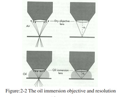
Because of refractive index the light passing from the glass into air makes the light to bend. The light passing from glass through oil does not bend much because the oil has similar refractive index to that of a glass.
The immersion oil with a refractive index of about 1.5 in-creases the numerical aperture and increases the resolving power of the microscope.
Related Topics