Chapter: Ophthalmology: Retina
Retina: Electrophysiologic Examination Methods
Electrophysiologic Examination Methods
(electroretinogram, electro-oculogram, and
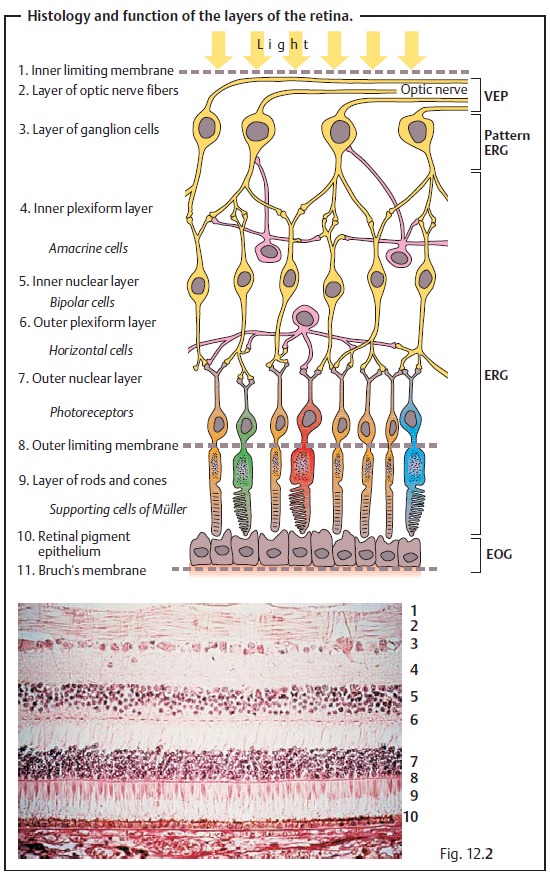
visual evoked potentials; see Fig. 12.2a)
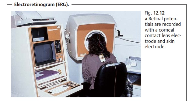
Electroretinogram (ERG):
This examination method uses electrodes to rec-ord the
electrical response of the retina to
flashes of light (Fig. 12.12a).
Photopic (light-adapted) and scotopic (dark-adapted) electroretinograms are
obtain-ed. The electroretinogram (ERG) consists of a negative A wave indicating
the response of the photoreceptors and a positive B wave primarily indicating
the response of the bipolar cells and the supporting cells of MĂĽller (Fig. 12.12b). A flicker ERG (repeated flashes) isolates pure cone response; a pattern ERG (such as a checkerboard)
and oscillating potentials can be used to evaluate the inner layers of the
retina. The ERG represents a summation
response of the ret-ina. A focal ERG
can record the response of isolated areas of the retina.
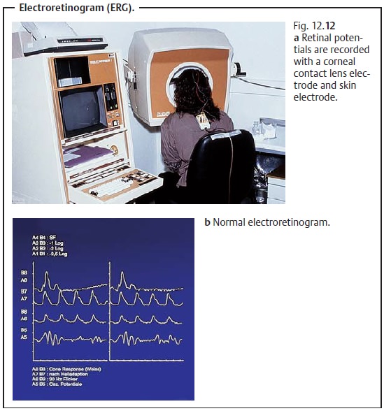
The classic indication for an
electroretinogram is retinitis pigmentosa with early loss of scotopic and
photopic potentials.
Electro-oculogram (EOG):
The electro-oculogram detectsabnormal changesin the retinal pigment epithelium such as macular vitelliform dystrophy. Thisexamination method utilizes the dipole of the eye in which the cornea forms the positive pole and the retinal pigment epithelium the negative pole. The standing potential across cornea and retina in comparison to the cornea is measured indirectly with two temporal electrodes (Fig. 12.13). During the measuring process, the patient performs regular eye movements by alter-nately focusing on two lights. The standing potential is normally higher in the light-adapted eye than in the dark-adapted eye.
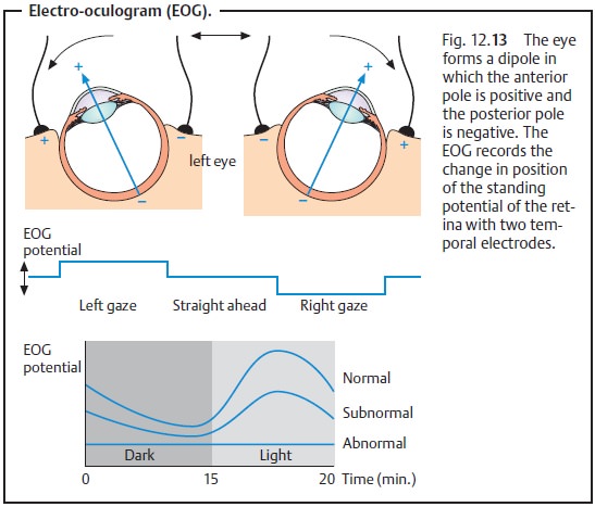
The ratio of light-adapted potential to dark-adapted potential (Arden ratio) is obtained to evaluate
the eye; this ratio is normally greater than 1.8. The ratio will be decreased
in the presence of abnormal changes. The typical indication for an
electro-oculogram is macular vitelliform dystrophy (Best’s vitelliform
dystrophy) with a significantly decreased Arden ratio.
Visual evoked potential (VEP):
This examination is
used to diagnose dam-age along the visual pathway. The VEP is not a specific
examination of the ret-ina such as an electroretinogram or electro-oculogram.
Related Topics