Chapter: Clinical Anesthesiology: Regional Anesthesia & Pain Management: Peripheral Nerve Blocks
Upper Extremity Peripheral Nerve Blocks: Brachial Plexus Anatomy

UPPER EXTREMITY PERIPHERAL NERVE BLOCKS
Brachial Plexus Anatomy
The brachial plexus is formed by the union of the anterior primary
divisions (ventral rami) of the fifth through the eighth cervical nerves and
the first thoracic nerves. Contributions from C4 and T2 are often minor or
absent. As the nerve roots leave the intervertebral foramina, they con-verge,
forming trunks, divisions, cords, branches, and then finally terminal nerves.
The three distinct trunks formed between the anterior and middle scalene
muscles are termed superior, middle, and inferior based on their vertical
orientation. As the trunks pass over the lateral border of the first rib

and under the clavicle, each trunk divides
into anterior and posterior divisions. As the brachial plexus emerges below the
clavicle, the fibers com-bine again to form three cords that are named
according to their relationship to the axillary artery: lateral, medial, and
posterior. At the lateral border of the pectoralis minor muscle, each cord
gives off a large branch before ending as a major terminal nerve. The lateral
cord gives off the lateral branch of the median nerve and terminates as the
musculocutaneous nerve; the medial cord gives off the medial branch of the
median nerve and termi-nates as the ulnar nerve; and the posterior cord gives
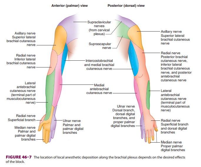
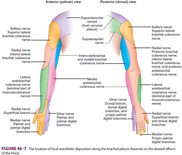
off the axillary nerve and terminates as the radial nerve. Local anesthetic may
be depos-ited at any point along the brachial plexus,depending on the desired block
effects (Figure 46–7): interscalene for shoulder and
proxi-mal humerus surgical procedures; and supracla-vicular, infraclavicular,
and axillary for surgeries distal to the mid-humerus.
Related Topics