Chapter: Essentials of Anatomy and Physiology: Nervous System
Spinal Nerves
SPINAL NERVES
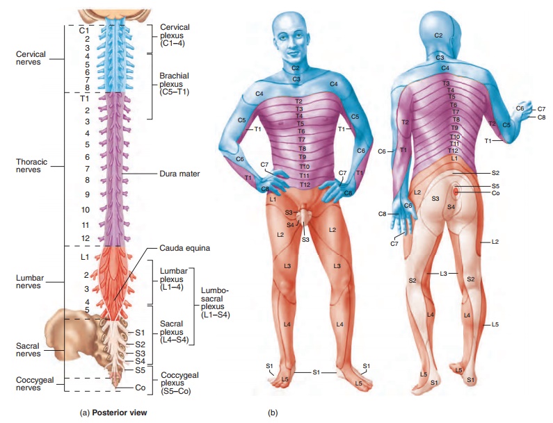
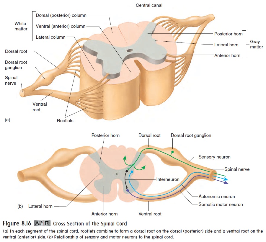
The spinal nerves arise along the spinal cord from the union of the dorsal roots and ventral roots (see figure 8.16). All the spinal nerves contain axons of both sensory and somatic motor neurons and thus are called mixed nerves. Some spinal nerves also contain parasympathetic or sympathetic axons. Most of the spinal nerves exit the vertebral column between adjacent vertebrae. Spinal nerves are categorized by the region of the vertebral column from which they emerge—cervical (C), thoracic (T), lumbar (L), sacral (S),
and coccygeal (Co). The spinal nerves are also numbered (start-ing superiorly) according to their order within that region. The 31 pairs of spinal nerves are therefore C1 through C8, T1 through T12, L1 through L5, S1 through S5, and Co (figure 8.20a).
The nerves arising from each region of the spinal cord and vertebral column supply specific regions of the body. A derma-tome is the area of skin supplied with sensory innervation by apair of spinal nerves. Each of the spinal nerves except C1 has a specific cutaneous sensory distribution. Figure 8.20b illustrates the dermatomal (der-m̆a -t̄o ′ m̆a l) map for the sensory cutaneous distribution of the spinal nerves.
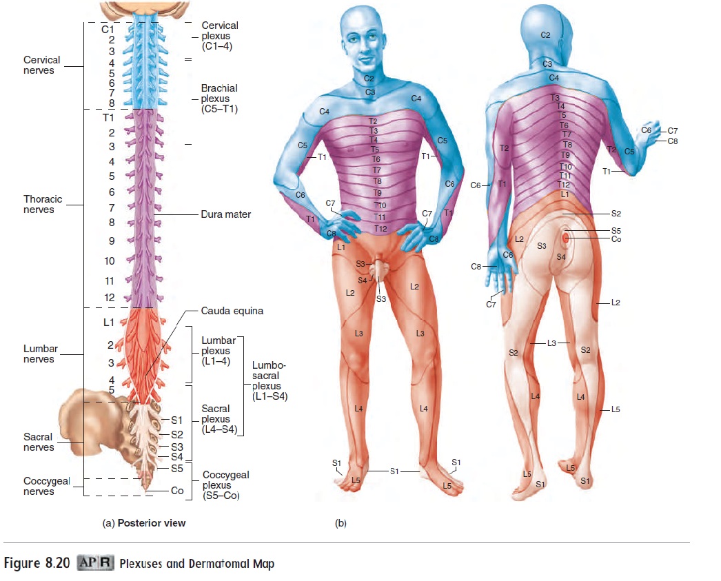
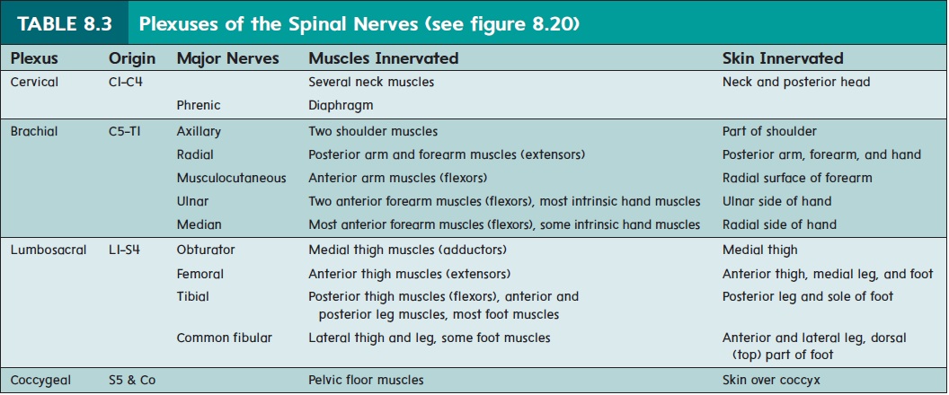
Most of the spinal nerves are organized into three major plexuses (plek′sŭs-ēz; braids) where neurons of several spinalnerves come together and intermingle. This reorganizes the neu-rons so that branches of nerves extending from each plexus contain neurons from different spinal segments. The three major plexuses are the cervical plexus, the brachial plexus, and the lumbosacral plexus (table 8.3). The major nerves of the neck and limbs are branches of these plexuses. Spinal nerves T2 through T11 do not join a plexus. Instead, these nerves extend around the thorax between the ribs, giving off branches to muscles and skin. Motor nerve fibers derived from plexuses innervate skeletal muscles, and sensory nerve fibers in those plexuses supply sensory innervation to the skin overlying those muscles (table 8.3). In addition to the major plexuses, the small coccygeal plexus supplies motor inner-vation to the muscles of the pelvic floor and sensory cutaneous innervation to the skin over the coccyx (figure 8.20b).
Cervical Plexus
The cervical plexus originates from spinal nerves C1 to C4. Branches from this plexus innervate several of the muscles attached to the hyoid bone, as well as the skin of the neck and posterior portion of the head. One of the most important branches of the cervical plexus is the phrenic nerve, which innervates the diaphragm. Contraction of the diaphragm is largely responsible for our ability to breathe .
Brachial Plexus
The brachial plexus originates from spinal nerves C5 to T1. Five major nerves emerge from the brachial plexus to supply the upper limb and shoulder. The axillary nerve innervates two shoulder muscles and the skin over part of the shoulder. The radial nerve innervates all the muscles in the posterior arm and forearm as well as the skin over the posterior surface of the arm, forearm, and hand. Themusculocutaneous (m̆u s′ k̄u -l̄o -k̄u -t̄a ′ n̄e -̆u s; muscle + skin) nerve innervates the anterior muscles of the arm and the skin over the radial surface of the forearm. The ulnar nerve inner-vates two anterior forearm muscles and most of the intrinsic hand muscles. It also innervates the skin over the ulnar side of the hand. The ulnar nerve can be easily damaged where it passes posterior to the medial side of the elbow. The ulnar nerve at this location is called the “funny bone.” The median nerve innervates most of the anterior forearm muscles and some of the intrinsic hand muscles. It also innervates the skin over the radial side of the hand.
Lumbosacral Plexus
The lumbosacral (l̆u m′ b̄o -s̄a ′ kr̆a l) plexus originates from spinal nerves L1 to S4. Four major nerves exit the plexus to supply the lower limb. The obturator (ob′ t̄u -r̄a -t̆o r) nerve innervates the muscles of the medial thigh and the skin over the same region. The femoral nerve innervates the anterior thigh muscles and the skinover the anterior thigh and medial side of the leg. The tibial nerve innervates the posterior thigh muscles, the anterior and posterior leg muscles, and most of the intrinsic foot muscles. It also innervates the skin over the sole of the foot. The common fibular (fib′ ̄u -l̆a r) nerve innervates the muscles of the lateral thigh and leg and someintrinsic foot muscles. It also innervates the skin over the anterior and lateral leg and the dorsal surface (top) of the foot. The tibial and common fibular nerves are bound together within a connective tis-sue sheath and together are called the sciatic (s̄ı -at′ ik) nerve.
Related Topics