Chapter: Essentials of Anatomy and Physiology: Nervous System
Autonomic Nervous System
AUTONOMIC NERVOUS SYSTEM
The autonomic nervous system (ANS) comprises motor neurons that carry action potentials from the CNS to the periphery. The autonomic neurons innervate smooth muscle, cardiac muscle, and glands. Autonomic functions are largely controlled unconsciously.
Axons from autonomic neurons do not extend all the way from the CNS to target tissues. This is in contrast to somatic motor neurons, which extend axons from the CNS to skeletal muscle. In the autonomic nervous system, two neurons in series extend from
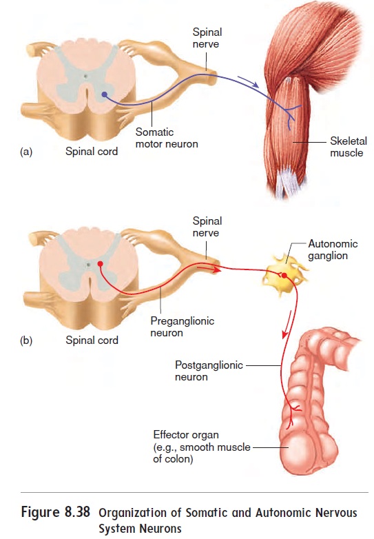
The first is called the preganglionicneuron; the second is the postganglionic neuron (figure 8.38).The neurons are so named because preganglionic neurons synapse with postganglionic neurons in autonomic ganglia outside the CNS. An exception is the preganglionic neuron that extends to the adrenal gland. There, the postganglionic neurons are actually the hormone-secreting cells of the adrenal medulla.
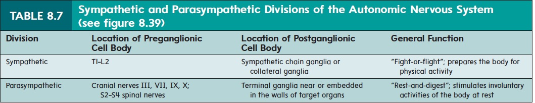
The autonomic nervous system is composed of the sympa-thetic (sim-pă-thet′ik;sympatheo,to feel with+pathos,suf-fering) division and the parasympathetic (par-ă-sim-pa-thet′ ik; para, alongside of)division(table 8.7 and figure 8.39). Increasedactivity in sympathetic neurons generally prepares the individual for physical activity, whereas parasympathetic stimulation gener-ally activates involuntary functions, such as digestion, that are normally associated with the body at rest.
Anatomy Of The Sympathetic Division
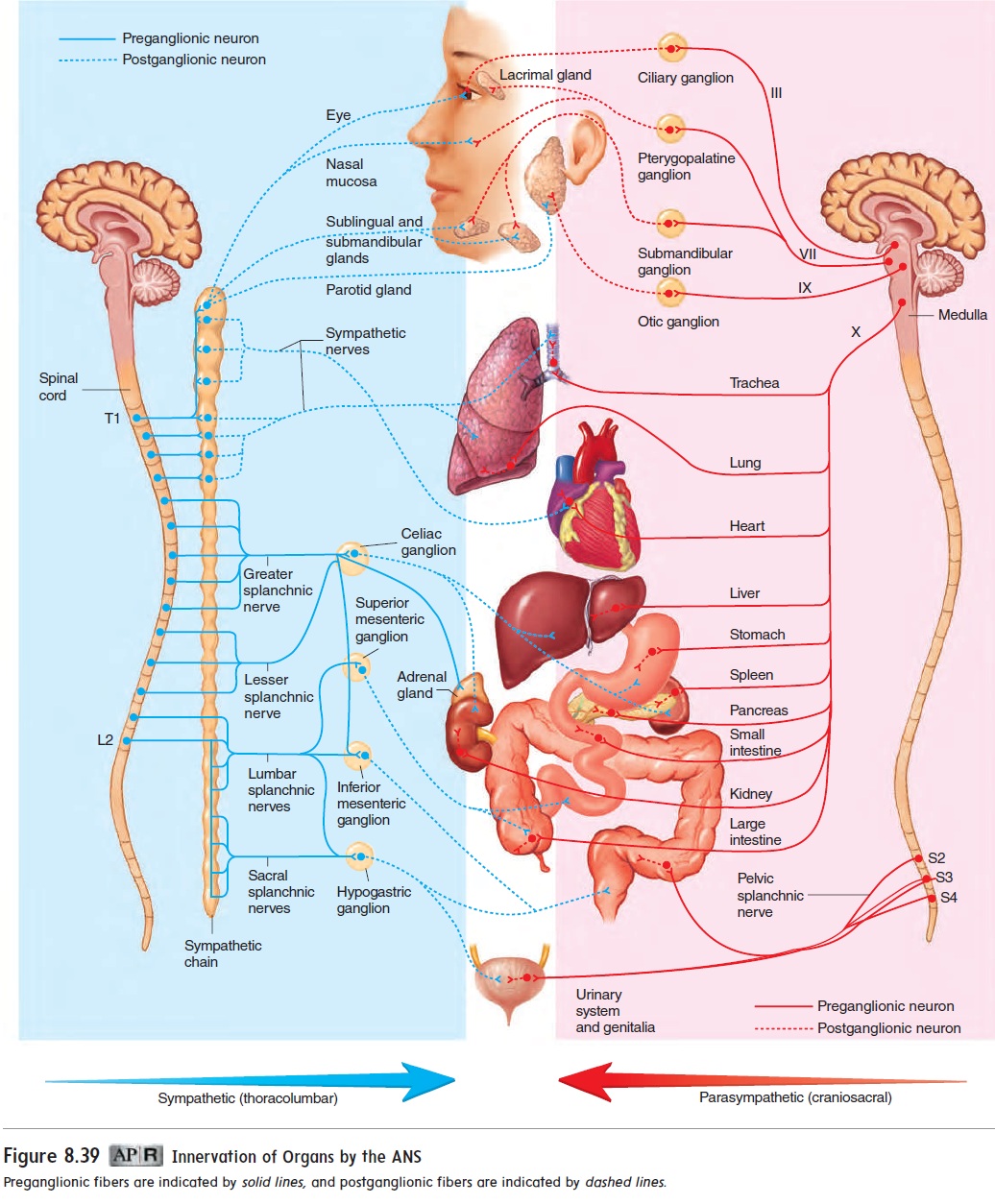
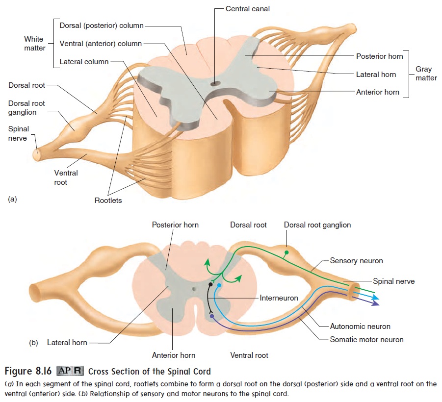
Cell bodies of sympathetic preganglionic neurons are in the lat-eral horn of the spinal cord gray matter (see figure 8.16) between the first thoracic (T1) and the second lumbar (L2) segments. The axons of the preganglionic neurons exit through ventral roots and project to either sympathetic chain ganglia or collat-eral ganglia (figure 8.39). The sympathetic chain ganglia are connected to one another and are so named because they form a chain along both sides of the spinal cord. Some preganglionic fibers synapse with postganglionic fibers in the sympathetic chain ganglia. The postganglionic axons form sympathetic nerves that innervate structures of the thoracic cavity. The axons of those preganglionic fibers that do not synapse in the sympathetic chain ganglia form splanchnic nerves that extend to collateral ganglia. Collateral ganglia are located nearer target organs and consist ofthe celiac, superior mesenteric, and inferior mesenteric ganglia. Preganglionic neurons synapse with postganglionic neurons in the collateral ganglia. Postganglionic neurons in the collateral ganglia project to target tissues in the abdominal and pelvic regions.
Anatomy Of The Parasympathetic Division
Preganglionic cell bodies of the parasympathetic division are located either within brainstem nuclei of the oculomotor nerve (III), facial nerve (VII), glossopharyngeal nerve (IX), or vagus nerve (X) or within the lateral part of the central gray matter of the spinal cord in the regions that give rise to spinal nerves S2 through S4.
Axons of the preganglionic neurons extend through spinal nerves or cranial nerves to terminal ganglia either located near effector organs in the head (figure 8.39) or embedded in the walls of effector organs in the thorax, abdomen, and pelvis.
The axons of the postganglionic neurons extend a relatively short distance from the terminal ganglia to the target organ. Most of the thoracic and abdominal organs are supplied by preganglionic neurons of the vagus nerve extending from the brainstem. The vagus nerve branches to provide parasympathetic innervation to the heart, the lungs, the liver, and the stomach and other digestive organs.
Autonomic Neurotransmitters
All preganglionic neurons of both the sympathetic and the para-sympathetic divisions and all the postganglionic neurons of the parasympathetic division secrete the neurotransmitter acetylcho-line. Most postganglionic neurons of the sympathetic divisionsecrete norepinephrine. Many body functions can be stimulated or inhibited by drugs that either mimic these neurotransmitters or prevent the neurotransmitters from activating their target tissues.
Functions of The Autonomic Nervous System
The sympathetic nervous system prepares a person for physical activity (table 8.8). These actions include increasing heart rate and blood pressure, dilating respiratory passageways to increase airflow, and stimulating the release of glucose from the liver for energy. At the same time, it inhibits digestive activities. In this way, the sympa-thetic division decreases the activity of organs not essential for the maintenance of physical activity and shunts blood and nutrients to structures that are active during exercise. In addition, excess heat is removed by vasodilation of the vessels near the skin and increased perspiration. The sympathetic division is sometimes referred to as the fight-or-flight system because it prepares the person either to stand and face a threat or to leave the situation as quickly as possible.
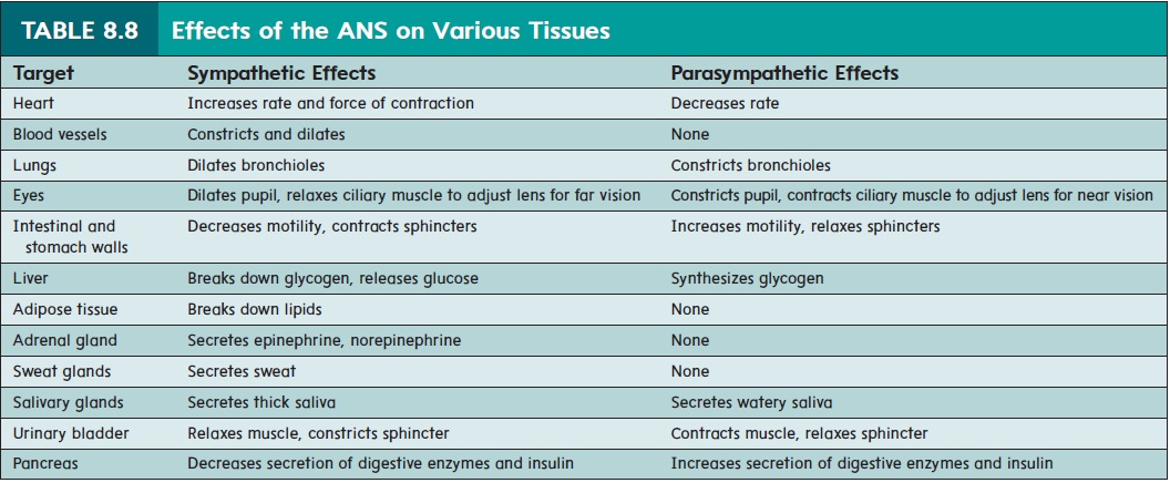
The parasympathetic division (rest-and-digest) of the auto-nomic nervous system is generally consistent with resting condi-tions (table 8.8). Increased activity of the parasympathetic division stimulates involuntary activities, such as digestion, defecation, and urination. The actions of the parasympathetic division in the diges-tive system illustrate how the ANS can coordinate the activities of multiple organs. Parasympathetic activity causes the release of digestive enzymes from the pancreas and contractions to mix the enzymes with food in the small intestine and move the material through the digestive tract. This cooperativity enhances the diges-tion and absorption of food. At the same time, parasympathetic stimulation lowers heart rate, which lowers blood pressure, and constricts air passageways, which decreases airflow.
The sympathetic and parasympathetic divisions can each pro-duce both stimulatory and inhibitory effects, depending on the tar-get tissue. For example, the sympathetic division stimulates smooth muscle contraction in blood vessel walls and inhibits smooth mus-cle contraction in lung airway walls. Likewise, the parasympathetic division stimulates contraction of the urinary bladder and inhibits contraction of the heart.
Most organs that receive autonomic neurons are innervated by both the parasympathetic and the sympathetic division. In most cases, the influences of the two autonomic divisions are opposite. For example, sympathetic stimulation of the heart causes an increase in heart rate, whereas parasympathetic stimulation causes a decrease in heart rate. When both divisions innervate a single organ, the sympathetic division tends to play a major role during physical activity or stress, whereas the parasympathetic division has more influence during resting conditions. Despite the general opposing actions of the two divisions, in some situa-tions, both divisions can act together to coordinate the activity of multiple targets. For example, in males, the parasympathetic divi-sion initiates erection of the penis, and the sympathetic division stimulates the release of secretions and helps initiate ejaculation. Not all organs receive dual innervation. Sweat glands and blood vessels are innervated by sympathetic neurons almost exclusively, whereas the smooth muscles associated with the lens of the eye are innervated primarily by parasympathetic neurons.
Related Topics