Chapter: Biochemical Pharmacology : Drugs that act on sodium and potassium channels
Some aspects of neurophysiology relevant to pharmacology
Some aspects of
neurophysiology relevant to pharmacology
This chapter presents some
basic facts from neurophysiol-ogy that will be needed in subsequent chapters on
the phar-macology of the nervous system.
The nervous system can be
divided according to different categories:
1. Central versus peripheral. The central nervous
system comprises the brain and the spinal cord, which together are protected
from the periphery by the blood brain bar-rier.
2. Somatic versus autonomic. The somatic nervous
system comprises functions that are conscious – conscious sen-sations such as
touch, temperature, pain etc., and vol-untary movements. Conversely, the
autonomic nervous system deals with unconscious sensory input such as blood
pressure, blood oxygen and carbon dioxide lev-els1, and the likewise
unconscious regulatory responses to it.
The two above distinctions
are `orthogonal', which means that we find autonomic and somatic parts in both
the cen-tral and the peripheral nervous system. Among the four re-sulting
categories, the peripheral autonomic system has a prominent role as a drug
target, for the following reasons:
• It is responsible for the functional regulation
of interior organs and physiological parameters (such as heart rate and blood
pressure) that are relevant to disease.
• It is readily accessible, i.e. not protected by
the blood brain barrier.
• As with all of the peripheral nervous system,
its orga-nization is comparatively simple, at least more so than that of the
brain is, and selective drug action is more fea-sible.
The relative simplicity of
the peripheral nervous system also explains why drug actions occurring there
usually are understood in a more definite way. With centrally act-ing drugs,
there often is a vagueness of understanding that makes them less compelling as
examples for an introducto-ry class – although in clinical practice they are
very impor-tant, of course.
Selective drug action on the
nervous system is feasible to the extent that different subsystems use specific
transmitters and receptors. Before we take a look at these, we will first
review some general aspects of nerve cell structure and function.
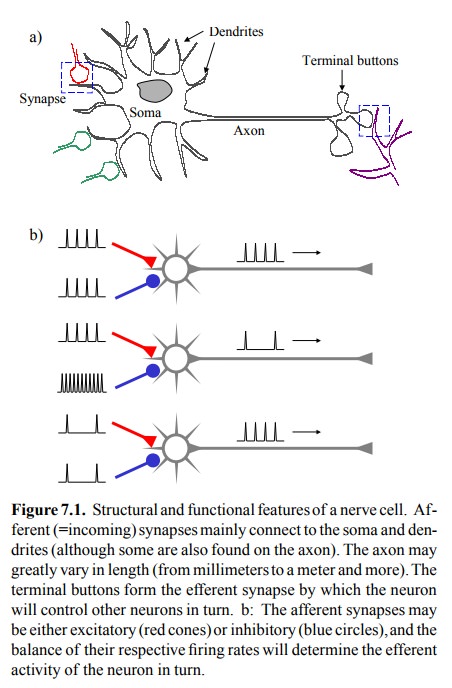
Figure 7.1a shows the
schematic of a nerve cell (neuron). The soma (greek: body) contains the nucleus
and the bulk of the biosynthetic apparatus (ER etc.) It branches out into
multiple dendrites (greek dendron = tree) and a single, usu-ally longer axon
that may branch in turn to reach multiple target cells. Afferent synapses are mostly located at the dendrites and the soma;
they receive signals from other neu-rons. The input from all dendritic synapses
is summed up and controls the efferent
activity of the neuron. The latter consists in the generation of action
potentials. The effer-ent action potentials travel swiftly down the axon to the
ef ferent synapses, where they will trigger the release of neu-rotransmitters
from the terminal buttons. In this way, they will influence the activity of the
next nerve or other ex-citable cell. All action potentials will be of similar
ampli-tude and duration; it is the repetition rate that represents the level of
activity (Figure 7.1b).

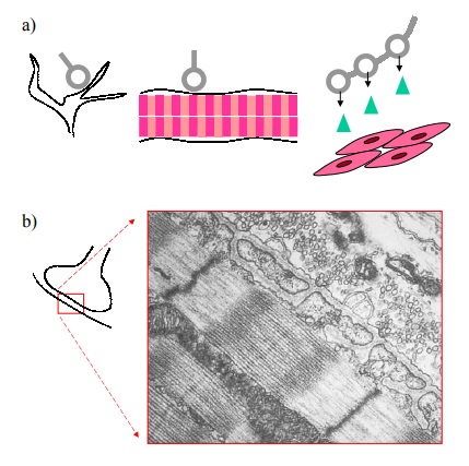
In all synapses, the
presynaptic cell will always be a neu-ron2. Postsynaptic cells can
be either neurons, striated or smooth muscle cells, or gland cells (Figure 7.2a).
In the case of skeletal muscle, the presynaptic neuron will be part of the
somatic nervous system. In contrast, neurons that project to the heart muscle
will be part of the autonomic system – none of us can voluntarily change the
heartbeat. While in many synapses the presynaptic and postsynaptic membranes
are in close apposition, thus ensuring rapid ac-tion, this is not necessarily
the case in the effector synapses of the autonomic nervous system, which
frequently do not have extremely time-critical missions.


Figure 7.2b shows an electron
microscopic picture of a neu-romuscular synapse. You can see the very narrow
synaptic cleft, the numerous synaptic vesicles, and the actin / myosin
filaments in the postsynaptic muscle cell. No space is wast-ed in this very
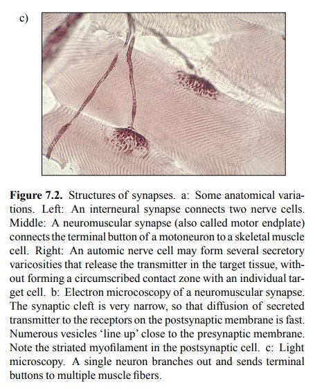
fast and efficient synapse. Figure 7.2c shows a lower power (light microscopic)
preparation of a nerve ending that branches toward multiple muscle cells. The
synaptic buttons are clearly visible. Note that a single nerve cell controls
several muscle fibers (up to several hundred, in fact). Synaptic excitation
will trigger an all-out action po-tential at the muscle cell membrane that will
be propagated swiftly along the entire expanse of the muscle fiber.
Related Topics