Chapter: Human Neuroanatomy(Fundamental and Clinical): Introduction to Neuroanatomy
Formation of neurons and neuroglial cells
Formation of neurons and neuroglial cells
The neurons and many neuroglial cells are formed in the neural tube.
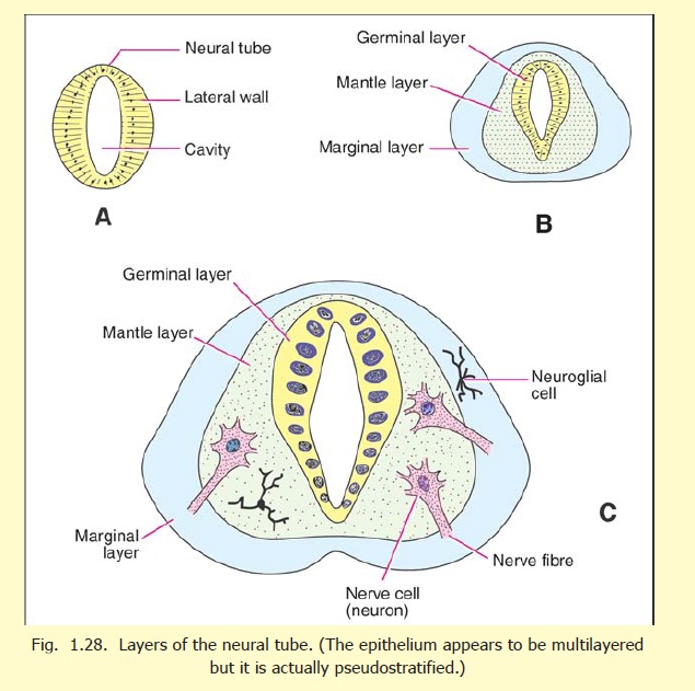
The neural tube is at first lined by a single layer of cells, (Fig. 1.28 A). These proliferate to form several layers (Fig. 1.28 B). Nearest the lumen of the tube is the matrix cell layer (also called primitive ependymal or germinal layer). The cells of this layer give rise to nerve cells, to neuroglial cells, and also to more germinal cells. Next comes the mantle layer in which are seen the developing nerve cells, and neuroglial cells. The outermost layer, termed the marginal zone, contains no nerve cells. It consists of a reticulum formed by protoplasmic processes of developing neuroglial cells (spongioblasts). It provides a framework into which the processes of nerve cells developing in the mantle layer can grow. (According to recent investigations the wall of the neural tube consists of only one layer of elongated cells. The multilayered appearance is produced by nuclei being placed at different levels as in a pseudostratified epithelium).
The stages in the formation of a nerve cell are as follows:
1. One of the germinal cells passes from the germinal layer to the mantle layer and becomes anapolar neuroblast (Fig. 1.29 A).
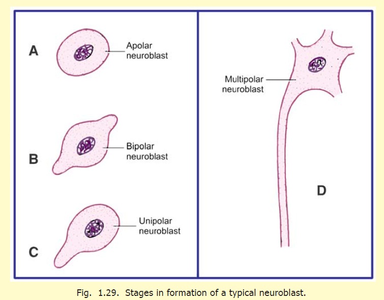
2. Two processes develop and convert the apolar neuroblast to a bipolar neuroblast (Fig. 1.29 B).
3. One of the processes of the neuroblast disappears, and it can now be called a unipolarneuroblast (Fig. 1.29 C).
4. The process of the cell that does not disappear now elongates, and on the side opposite to it numerous smaller processes form. At this stage the cell is called a multipolar neuroblast (Fig. 1.29 D).
5. The main process of the multipolar neuroblast now grows into the marginal layer, and becomes the axon of the nerve cell (Fig. 1.29 B). The axon can grow to a considerable length. It may either remain within the central nervous system, or may grow out of it as an efferent nerve fibre of a peripheral nerve. At its destination it establishes connections, either with the cell bodies and dendrites of other neurons, or with an effector organ (e.g. muscle).
6. The smaller processes of the neuroblast are the dendrites. These ramify and establish connections with other nerve cells.
7. At first the cytoplasm of the nerve cell is homogeneous. Later Nissl’s granules make their appearance. After their formation, neurons lose the ability to divide.
Neuroglial cells are also formed from germinal cells of the ependymal layer. These cells (glioblasts) migrate to the mantle and marginal zones as medulloblasts (also called spongioblasts), which differentiate either into astroblasts, and subsequently intoastrocytes, or into oligodendroblasts and then into oligodendrocytes. There is a third type of neuroglial cell called microglia. This type does not develop from the cells of the neural tube, but migrates into it along with blood vessels. These cells are believed to be of mesodermal origin.
We have seen above that the ependymal (or neuroepithelial) cells give rise both to neuroblasts and to neuroglia. However, these two cell types are not formed simultaneously. The neuroblasts are formed first. Neuroglial cells are formed after the differentiation of neuroblasts is completed.
The formation of the myelin sheath by Schwann cells and oligodendrocytes has already been explained. Nerve fibres in different parts of the brain, and spinal cord, become myelinated at different stages of development. The process begins during the fourth month of intrauterine life, but is not completed until the child is two to three years old. Nerve fibres become fully functional only after they have acquired their myelin sheaths.
The blood vessels of the brain, and their surrounding connective tissue, are not derived from the neural tube. These are mesodermal in origin and invade the developing brain and spinal cord from the surrounding mesoderm.
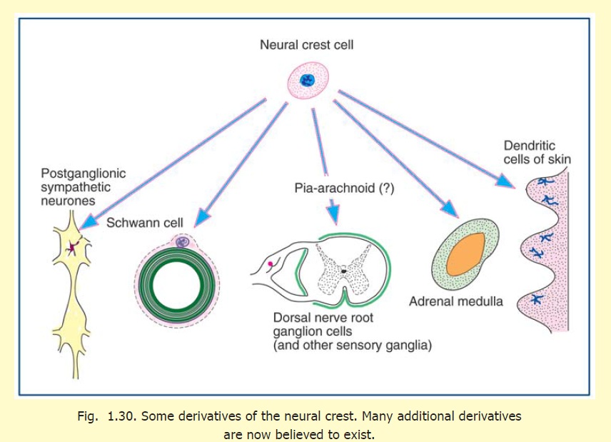
The development of the pia mater and the arachnoid mater (leptomeninges) is not definitely understood. According to some workers, these are derived from the neural crest. The dura mater develops from the mesoderm surrounding the neural tube.
There can be errors in the closure of the neural tube. This leads to congenital anomalies line anencephaly and rachischisis.
Related Topics