Chapter: Human Neuroanatomy(Fundamental and Clinical): Introduction to Neuroanatomy
Basic Structure of Peripheral Nerve Fibres
Basic Structure of Peripheral Nerve Fibres
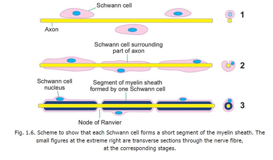
Each nerve fibre has a central core formed by the axon. This core is called the axis cylinder. The plasma membrane surrounding the axis cylinder is the axolemma. The axis cylinder is surrounded by a myelin sheath. This sheath is in the form of short segments that are separated at short intervals called the nodes of Ranvier (Fig. 1.6). The part of the nerve fibre between two consecutive nodes is the internode.
Each segment of the myelin sheath is formed by one Schwann cell. Outside the myelin sheath there is a thin layer of Schwann cell cytoplasm. This layer of cytoplasm is called the neurilemma. The method of formation of these sheaths has been described.

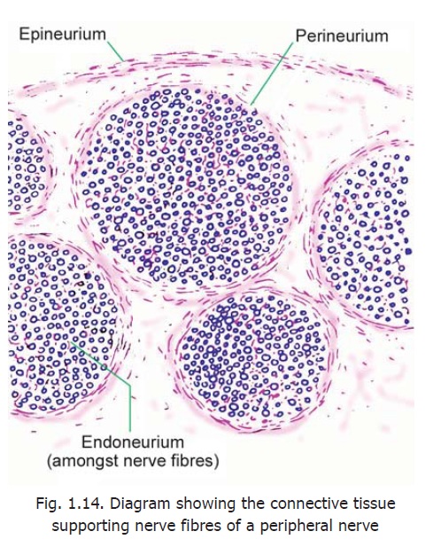
Each nerve fibre is surrounded by endoneurium (Fig. 1.14). This is a layer ofconnective tissue The endoneurium holds adjoining nerve fibres together and facilitates their aggregation to form bundles or fasciculi. Apart from collagen fibres the endoneurium contains fibroblasts, Schwann cells, endothelial cells and macrophages.

Each fasciculus is surrounded by the perineurium, which is a thicker layer ofconnective tissue. The perineurium is made up of layers of flattened cells separated by layers of collagen fibres. The perineurium probably controls diffusion of substances in and out of axons.
A very thin nerve may consist of a single fasciculus, but usually a nerve is made up of several fasciculi. The fasciculi are held together by the epineurium. This is a fairly dense layer of connective tissue that surrounds the entire nerve.
The epineurium contains fat that cushions nerve fibres. Loss of this fat in bedridden patients can lead to pressure on nerve fibres and paralysis.
Blood vessels to a nerve travel through the connective tissue that surrounds it. Severe reduction in blood supply can lead toischaemic neuritis and pain.
Related Topics