Chapter: Essentials of Anatomy and Physiology: Nervous System
Motor Functions: Motor Areas Of The Cerebral Cortex, Descending tracts, Cerebellum
Motor Functions
The motor system of the brain and spinal cord is responsible for maintaining the body’s posture and balance, as well as moving the trunk, head, limbs, tongue, and eyes and communicating through facial expressions and speech. Reflexes mediated through the spinal cord and brainstem are responsible for some body movements. These are called involuntary movements because they occur without conscious thought. Voluntary movements, on the other hand, are consciously activated to achieve a specific goal, such as walking or typing. Although consciously activated, the details of most voluntary movements occur automatically. For example, once you start walking, you don’t have to think about the moment-to-moment control of every muscle because neural circuits in the reticular formation and spinal cord automatically control your limbs. Once learned, complex tasks, such as typing, can be performed relatively automatically.
Voluntary movements result from the stimulation of upper and lower motor neurons. Upper motor neurons have cell bodies in the cerebral cortex. The axons of upper motor neurons form descending tracts that connect to lower motor neurons. Lowermotor neurons have cell bodies in the anterior horn of the spinalcord gray matter or in cranial nerve nuclei. Their axons leave the central nervous system and extend through spinal or cranial nerves to skeletal muscles..
Motor Areas Of The Cerebral Cortex
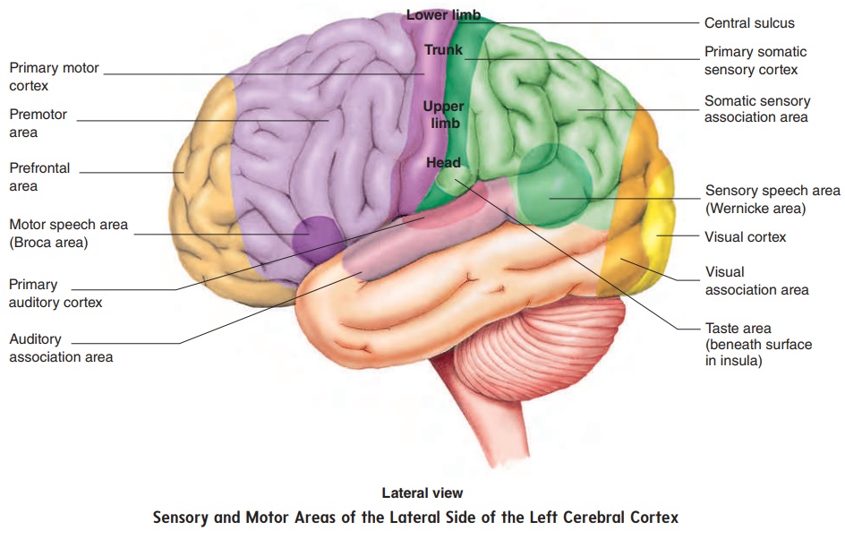
The primary motor cortex is located in the posterior portion of the frontal lobe, directly anterior to the central sulcus (figure 8.27). Action potentials initiated in this region control voluntary move-ments of skeletal muscles. Upper motor neuron axons project from specific regions of this cortex to specific parts of the body so that a topographic map of the body exists in the primary motor cortex, with the head inferior, analogous to the topographic map of the primary somatic sensory cortex (figure 8.27). The premotor area of the frontal lobe is where motor functions are organized before they are actually initiated in the primary motor cortex. For example, if a person decides to take a step, the neurons of the premotor area are first stimulated, and the determination is made there as to which muscles must contract, in what order, and to what degree. Action potentials are then passed to the upper motor neurons of the pri-mary motor cortex, which initiate each planned movement.
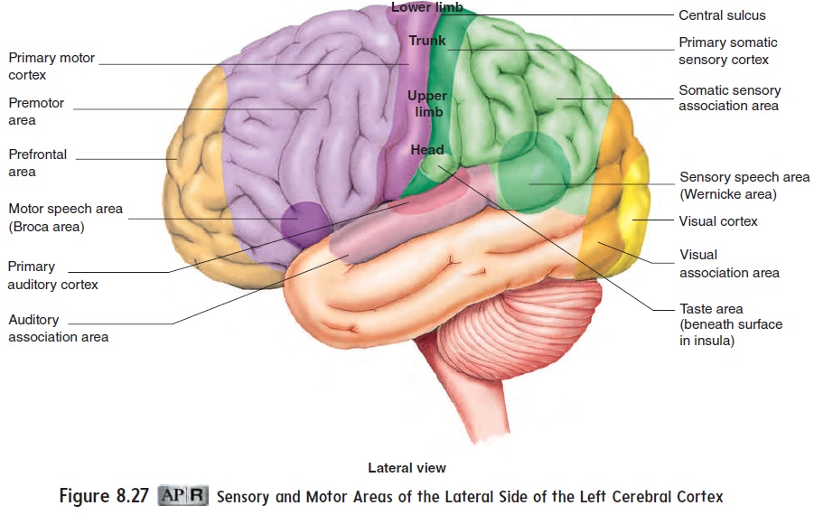
The motivation and foresight to plan and initiate movements occur in the anterior portion of the frontal lobes, called the pre-frontal area. This region of association cortex is well developedonly in primates, especially humans. It is involved in motivation and regulation of emotional behavior and mood. The large size of this area in humans may account for our emotional complexity and our relatively well-developed capacity to think ahead and feel motivated.
Descending tracts
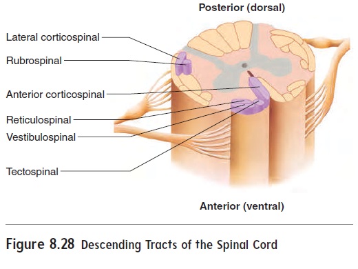
The names of the descending tracts are based on their origin and ter-mination (table 8.5 and figure 8.28). For example, the corticospinal tracts are so named because they begin in the cerebral cortex and terminate in the spinal cord. The corticospinal tracts are considered direct because they extend directly from upper motor neurons in thecerebral cortex to lower motor neurons in the spinal cord (a similar direct tract extends to lower motor neurons in the brainstem). Other tracts are named after the part of the brainstem from which they originate. Although they originate in the brainstem, these tracts are indirectly controlled by the cerebral cortex, basal nuclei, and cerebel-lum. These tracts are called indirect because no direct connection exists between the cortical and spinal neurons.
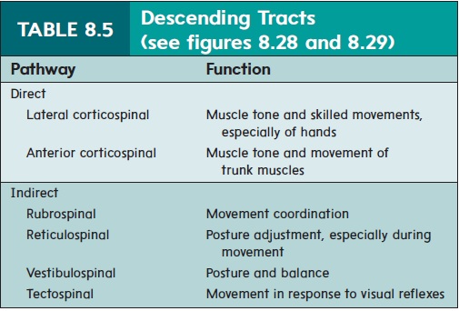
The descending tracts control different types of movements (table 8.5). Tracts in the lateral columns (see figure 8.16; figure 8.28) are most important in controlling goal-directed limb movements, such as reaching and manipulating. The lateral corticospinaltracts are especially important in controlling the speed and precisionof skilled movements of the hands. Tracts in the ventral columns, such as the reticulospinal tract, are most important for maintaining posture, balance, and limb position through their control of neck, trunk, and proximal limb muscles.
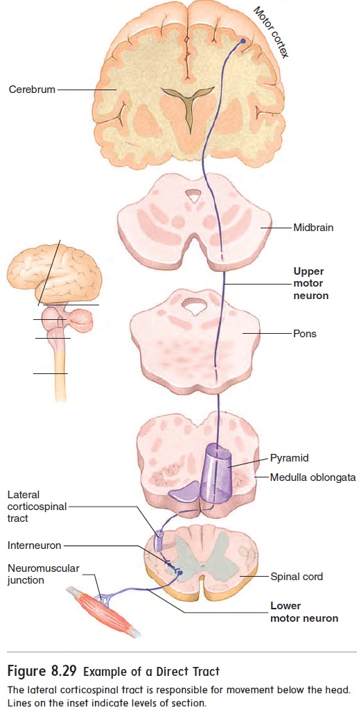
The lateral corticospinal tract serves as an example of how descending pathways function. It begins in the cerebral cortex and descends into the brainstem (figure 8.29). At the inferior end of the pyramids of the medulla oblongata, the axons cross over to the oppo-site side of the body and continue into the spinal cord. Crossover of axons in the brainstem or spinal cord to the opposite side of the body is typical of descending pathways. Thus, the left side of the brain controls skeletal muscles on the right side of the body, and vice versa. The upper motor neuron synapses with interneurons that then synapse with lower motor neurons in the brainstem or spinal cord. The axon of the lower motor neuron extends to the skeletal muscle fiber.
Basal nuclei
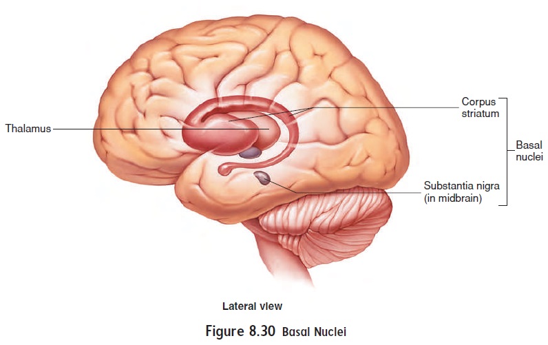
The basal nuclei are a group of functionally related nuclei (figure 8.30). Two primary nuclei are the corpus striatum (kōr′ -pŭs strı̄-ā′ tŭm), located deep within the cerebrum, and the substantianigra, a group of darkly pigmented cells in the midbrain.
The basal nuclei are important in planning, organizing, and coordinating motor movements and posture. Complex neural cir-cuits link the basal nuclei with each other, with the thalamus, and with the cerebral cortex. These connections form several feedback loops, some of which are stimulatory and others inhibitory. The stimulatory circuits facilitate muscle activity, especially at the
beginning of a voluntary movement, such as rising from a sitting position or beginning to walk. The inhibitory circuits facilitate the actions of the stimulatory circuits by inhibiting muscle activity in antagonist muscles. In addition, inhibitory circuits inhibit random movements of the trunk and limbs. Inhibitory circuits also decrease muscle tone when the body, limbs, and head are at rest. Disorders of the basal nuclei result in difficulty rising from a sitting position and difficulty initiating walking. People with basal nuclei disorders exhibit increased muscle tone and exaggerated, uncontrolled move-ments when they are at rest. A specific feature of some basal nuclei disorders is “resting tremor,” a slight shaking of the hands when a person is not performing a task. Parkinson disease, Huntington dis-ease, and cerebral palsy are basal nuclei disorders (see the Diseases and Disorders table).
Cerebellum
The cerebellum is attached by cerebellar peduncles to the brainstem (see figures 8.22 and 8.24b). The cerebellar cortex is composed of gray matter and has gyri and sulci, but the gyri are much smaller than those of the cerebrum. Internally, the cerebellum consists of gray nuclei and white nerve tracts. The cerebellum is involved in maintaining balance and muscle tone and in coordinating fine motor movement. If the cerebellum is damaged, muscle tone decreases, and fine motor movements become very clumsy.
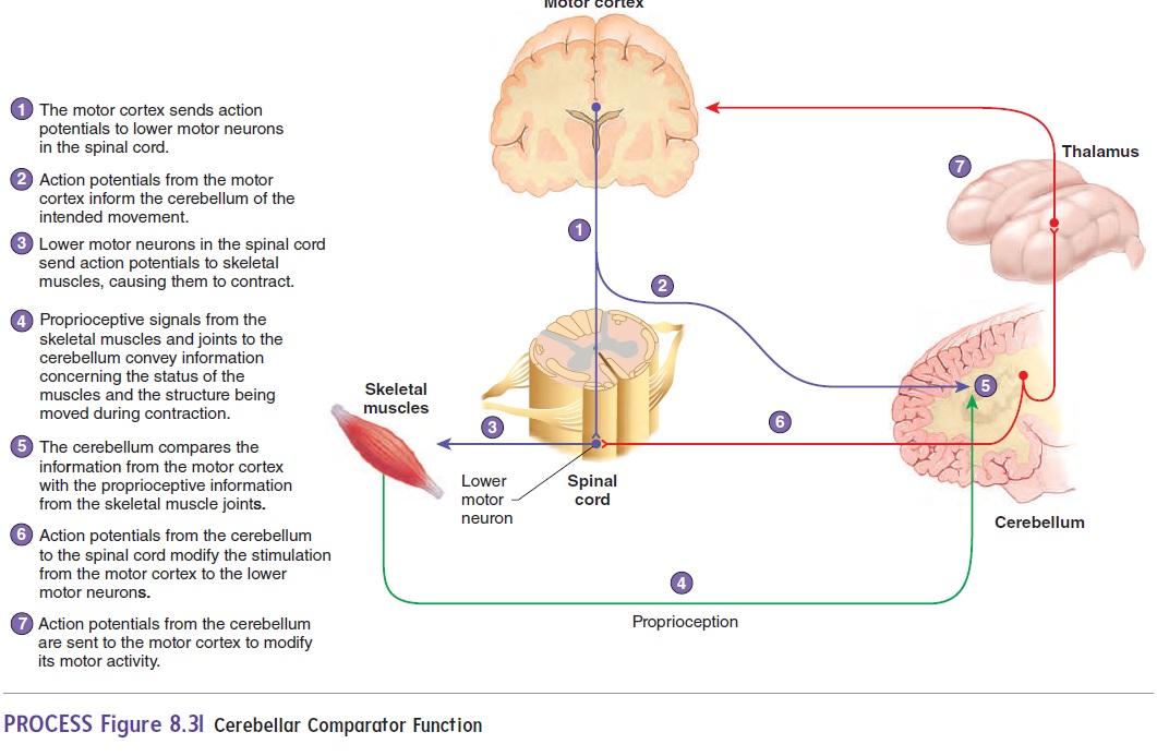
A major function of the cerebellum is that of a comparator (figure 8.31). A comparator is a sensing device that compares the data from two sources—in this case, the motor cortex and peripheral structures. Action potentials from the cerebral motor cortex descend into the spinal cord to initiate voluntary movements. Collateral branches are also sent from the motor cortex to the cer-ebellum, giving information representing the intended movement. In addition, simultaneously, reaching the cerebellum are action potentials fromproprioceptive (prō-prē-ō-sep′ tiv) neurons, which innervate joints, tendons, and muscles and provide information about the position of body parts. The cerebellum compares infor-mation about the intended movement from the motor cortex to sensory information from the moving structures. If a difference is detected, the cerebellum sends action potentials to motor neurons in the motor cortex and the spinal cord to correct the discrepancy. The result is smooth and coordinated movements. For example, if you close your eyes, the cerebellar comparator function allows you to touch your nose smoothly and easily with your finger. If the cer-ebellum is not functioning, your finger tends to overshoot the target. One effect of alcohol is to inhibit the function of the cerebellum.
Another function of the cerebellum involves participating with the cerebrum in learning motor skills, such as playing the piano. Once the cerebrum and cerebellum “learn” these skills, the special-ized movements can be accomplished smoothly and automatically.
Related Topics