Sexual Reproduction in Plants - Male Reproductive part - Androecium | 12th Botany : Chapter 1 : Asexual and Sexual Reproduction in Plants
Chapter: 12th Botany : Chapter 1 : Asexual and Sexual Reproduction in Plants
Male Reproductive part - Androecium
Pre-fertilization structure and events
The hormonal and
structural changes in plant lead to the differentiation and development of
floral primordium. The structures and events involved in pre-fertilization are
given below
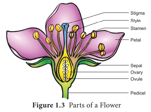
Male Reproductive part - Androecium
Androecium is made up of
stamens. Each stamen possesses an anther and a filament. Anther bears pollen
grains which represent the male gametophyte. In this chapter we shall discuss
the structure and development of anther in detail.
Development of anther:
A very young anther develops as a
homogenous mass of cells surrounded by an epidermis. During its development,
the anther assumes a four-lobed structure. In each lobe, a row or a few rows of
hypodermal cells becomes enlarged with conspicuous nuclei. This functions as
archesporium. The archesporial cells divide by periclinal divisions to form
primary parietal cells towards the epidermis and primary sporogenous cells
towards the inner side of the anther. The primary parietal cells undergo a
series of periclinal and anticlinal division and form 2-5 layers of anther
walls composed of endothecium, middle layers and tapetum, from periphery to
centre.
Microsporogenesis: The stages involved in the formation of haploid microspores from diploid microspore mother cell through meiosis is called Microsporogenesis. The primary sporogeneous cells directly, or may undergo a few mitotic divisions to form sporogenous tissue.
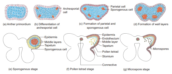
The last generation of
sporogenous tissue functions as microspore mother cells. Each microspore mother
cell divides meiotically to form a tetrad of four haploid microspores
(microspore tetrad). The microspore tetrad may be arranged in a tetrahedral,
decussate, linear, T shaped or isobilateral manner. Microspores soon separate
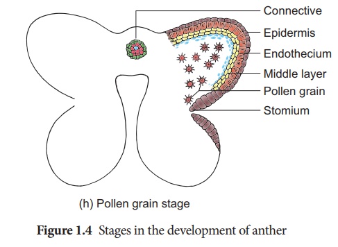
from one another and remain free in the anther locule and develop into pollen
grains. The stages in the development of microsporangia is given in Figure 1.4.
In some plants, all the microspores in a microsporangium remain held together
called pollinium. Example: Calotropis. Compound pollen grains are
found in Drosera and Drymis.
T.S. of Mature anther
Transverse section of
mature anther reveals the presence of anther cavity surrounded by an anther
wall. It is bilobed, each lobe having 2 theca (dithecous). A typical anther is
tetrasporangiate. The T.S. of Mature anther is given in Figure 1.5.
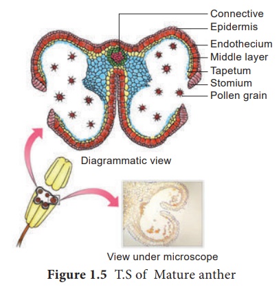
1. Anther wall
The mature anther wall
consists of the following layers a. Epidermis b. Endothecium c.
Middle layers d. Tapetum.
a. Epidermis: It is single layered
and protective in function. The cells undergo repeated anticlinal
divisions to cope up with the rapidly enlarging internal tissues.
b. Endothecium: It is generally a single layer of radially elongated cells found below the epidermis. The inner tangential wall develops bands (sometimes radial walls also) of α cellulose (sometimes also slightly lignified). The cells are hygroscopic. In the anthers of aquatic plants, saprophytes, cleistogamous flowers and extreme parasites endothecial differentiation is absent. The cells along the junction of the two sporangia of an anther lobe lack these thickenings. This region is called stomium. This region along with the hygroscopic nature of endothecium helps in the dehiscence of anther at maturity.
c. Middle layers: Two to three layers of
cells next to endothecium constitute middle layers. They are generally
ephemeral. They disintegrate or get crushed during maturity.
d. Tapetum: It is the innermost
layer of anther wall and attains its maximum development at the tetrad
stage of microsporogenesis. It is derived partly from the peripheral wall layer
and partly from the connective tissue of the anther lining the anther locule.
Thus, the tapetum is dual in origin. It nourishes the developing sporogenous
tissue, microspore mother cells and microspores. The cells of the tapetum may
remain uninucleate or may contain more than one nucleus or the nucleus may
become polyploid. It also contributes to the wall materials, sporopollenin, pollenkitt,
tryphine and number of proteins that control incompatibility reaction .Tapetum
also controls the fertility or sterility of the microspores or pollen grains.
There are two types of
tapetum based on its behaviour. They are:
Secretory tapetum (parietal/glandular/ cellular):
The tapetum retains the original position and cellular integrity and nourishes
the developing microspores.
Invasive tapetum (periplasmodial): The cells
loose their inner tangential and radial walls and the protoplast of all tapetal
cells coalesces to form a periplasmodium.
Functions of Tapetum:
·
It supplies nutrition to the developing microspores.
·
It contributes sporopollenin through ubisch bodies thus
plays an important role in pollen wall formation.
·
The pollenkitt material is contributed by tapetal cells and is
later transferred to the pollen surface.
Exine proteins
responsible for ‘rejection reaction’ of the stigma are present in
the cavities of the exine. These proteins are derived from tapetal
cells.
2. Anther Cavity : The anther cavity is
filled with microspores in young stages or with pollen grains at
maturity. The meiotic division of microspore mother cells gives rise to
microspores which are haploid in nature.
3. Connective: It is the column of
sterile tissue surrounded by the anther lobe. It possesses vascular
tissues. It also contributes to the inner tapetum.
Microspores and pollen grains
Microspores are the
immediate product of meiosis of the microspore mother cell whereas the pollen
grain is derived from the microspore. The microspores have protoplast
surrounded by a wall which is yet to be fully developed. The pollen protoplast
consists of dense cytoplasm with a centrally located nucleus. The wall is
differentiated into two layers, namely, inner layer called intine and
outer layer called exine. Intine is thin, uniform and is made up of
pectin, hemicellulose, cellulose and callose together with proteins. Exine is
thick and is made up of cellulose, sporopollenin and pollenkitt. The exine is
not uniform and is thin at certain areas. When these thin areas are small and
round it is called germ pores or when elongated it is called furrows. It is
associated with germination of pollen grains. The sporopollenin is generally
absent in germ pores.The surface of the exine is either smooth or sculptured in
various patterns (rod like, grooved, warty, punctuate etc.) The sculpturing
pattern is used in the plant identification and classification.
Shape of a pollen grain
varies from species to species. It may be globose, ellipsoid, fusiform, lobed,
angular or crescent shaped. The size of the pollen varies from 10 micrometers
in Myosotis to 200 micrometers in members of the family Cucurbitaceae
and Nyctaginaceae
The wall material
sporopollenin is contributed by both pollen cytoplasm and tapetum. It is
derived from carotenoids. It is resistant to physical and biological
decomposition. It helps to withstand high temperature and is resistant to
strong acid, alkali and enzyme action. Hence, it preserves the pollen for long
periods in fossil deposits, and it also protects pollen during its journey from
anther to stigma.
Pollenkitt is
contributed by the tapetum and coloured yellow or orange and is chiefly made of
carotenoids or flavonoids. It is an oily layer forming a thick viscous coating
over pollen surface. It attracts insects and protects damage from UV radiation.
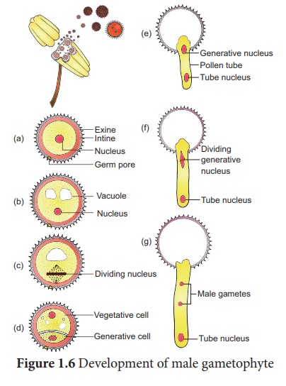
Development of Male gametophyte:
The microspore is the
first cell of the male gametophyte and is haploid. The development of male
gametophyte takes place while they are still in the microsporangium. The
nucleus of the microspore divides to form a vegetative and a generative
nucleus. A wall is laid around the generative nucleus resulting in the
formation of two unequal cells, a large irregular nucleus bearing with abundant
food reserve called vegetative cell and a small generative cell. At this 2
celled stage, the pollens are liberated from the anther. In some plants the
generative cell again undergoes a division to form two male gametes. In these
plants, the pollen is liberated at 3 celled stage. In 60% of the angiosperms
pollen is liberated in 2 celled stage. Further, the growth of the male
gametophyte occurs only if the pollen reaches the right stigma. The pollen on
reaching the stigma absorbs moisture and swells. The intine grows as pollen tube
through the germ pore. In case the pollen is liberated at 2 celled stage the
generative cell divides in the pollen into 2 male cells (sperms) after reaching
the stigma or in the pollen tube before reaching the embryo sac. The stages in
the development of male gametophyte is given in Figure 1.6.
Related Topics