Chapter: Orthopaedics
Articular Cartilage Defects
Articular Cartilage
Defects
Properties of Articular Cartilage
·
lacks blood supply and does not have innervation
or lymphatic drainage
·
varies in thickness from 2 mm to 4 mm and is
thickest at periphery of concave surfaces and central portions of convex
surfaces
·
composed of type 2 collagen, water, proteoglycans,
and chondrocytes
·
collagen provides resistance against tensile
stresses and transmits vertical loads
·
water and proteoglycans provide turgor and
elasticity and help to limit friction
·
chondrocytes synthesize the cartilage matrix and
control matrix turnover rate
Etiology
·
overt trauma or repeated minor trauma; most
commonly from sports injuries
·
early stage osteoarthritis
·
genetic degenerative diseases such as
osteochondritis dissecans
Clinical Features
·
very similar to symptoms of osteoarthritis (joint
line pain with possible effusion, etc.)
·
often have predisposing factors such as ligament
injury, malalignment of the joint (varus/ valgus), obesity, bone deficiency
(avascular necrosis, osteochondritis dissecans, ganglion bone cysts),
inflammatory arthropathy, and familial osteoarthropathy
·
may have symptoms of locking or catching related to
the torn/displaced cartilage
Investigations
·
arthroscopy to visualize focal pathology and guide
treatment strategy
·
MRI may also be used to visualize the defect
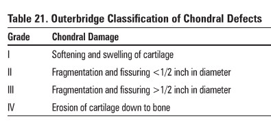
Table 21.
Outerbridge Classification of Chondral Detects
Treatment
·
arthroscopic lavage and debridement of the joint
·
marrow stimulation techniques (microfracture,
drilling, abrasion arthroplasty)
o
involves creating a site of bleeding where new
growth/healing can take place
·
osteochondral grafts; also known as the OATS
procedure or mosaicplasty
o
involves transferring osteochondral fragments from
non-weightbearing surface to area of defect
·
autologous chondrocyte: implantation (ACI)
o
currently only available in the U.S. and Europe
o
involves harvesting patient's cartilage, growing
it in culture: medium outside of the patient, then reinserting the newly
cultured chondrocytes back to fill the chondral defect
o
osteochondral allograft; only used in limited
circumstances when defect is very large
Related Topics