Chapter: 11th 12th std standard Home Science Maintain Basic Knowledge for family life Higher secondary school College
Ear : Physiology of Hearing
Physiology
of Hearing
Every
sound produces sound waves or disturbances in the air, which travel at about
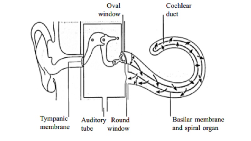
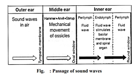
332 metres (1088) feet per second. The auricle, because of its shape,
concentrates the waves and directs them along the auditory meatus causing the
tympanic membrane to vibrate.
Tympanic
membrane vibrations are transmitted through the middle ear by movement of the
ossicles. At their medial end the footplate of the stapes rock to and fro in
the oval window, setting up fluid waves in the perilymph. These indent the
membranous labyrinth and the wave motion in the endolymph stimulates the
neuroepithelial cells of the organ of Corti. The nerve impulses produced pass
to the brain in the cochlear portion of the eighth cranial nerve (VIII). The
fluid wave is finally expended into the middle ear by vibration of the membrane
of the round window. This nerve, the vestibulocochlear nerve, transmits the
impulse to various nuclei in the pons varolii and midbrain. Some of the nerve
fibres pass to the hearing area in the cerebral cortex where sound is
perceived.
Semicircular
Canals
The semicircular canals have no auditory function although they are
closely associated with the cochlea. They provide information about the
position of the head in space, contributing to maintenance of equilibrium and
balance.
There are three semicircular canals, one lying in each of the three
planes of space. They are situated above and behind the vestibule of the inner
ear and open into it.
Structure of the semircular canals
The semicircular canals, like the cochlea are composed of an outer bony
wall and inner membranous tubes or ducts. The membranous ducts contain
endolymph and are separated from the bony wall by perilymph.
The utricle is a membranous
sac which is part of the vestibule and the three membranous ducts open into it
at their dilated ends, the ampullae.
The saccule is a part of the
vestibule and communicates with the utricle and the cochlea.
In the
walls of the utricle, saccule and ampullae there are fine specialized
epithelial cells with minute projections, called hair cells. Amongst the hair cells there are the minute nerve
endings of the vestibular part of the vestibulocochlear
nerve.
Functions of the
semicircular canal
The semicircular canals, utricle and saccule are concerned with balance.
Any change of position of the head causes movement in the perilymph and
endolymph which stimulates the nerve endings and the hair cells in the utricle,
saccule and ampullae. The resultant nerve impulses are transmitted by the vestibular
nerve to the cerebellum.
The cerebellum also receives nerve impulses from
the eyes and the muscles and joints. Impulses from these three sources are
coordinated and efferent nerve impulses pass to the cerebrum where position in
space is perceived, and to muscles to maintain posture and balance.
Related Topics