Chapter: 11th 12th std standard Home Science Maintain Basic Knowledge for family life Higher secondary school College
Structure of Ear
The Ear
The ear is the organ of hearing. It is supplied by the eighth cranial nerve, i.e., the cochlear part of the vestibulocochlear nerve which is
stimulated by vibrations caused by sound waves.
With the
exception of the auricle (pinna), the structures that form the ear are encased
within the temporal bone.
Structure
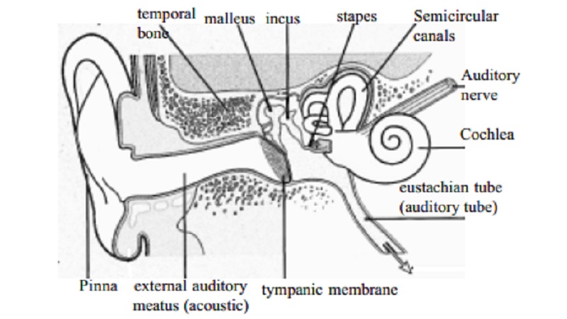
The ear is divided into three distinct parts.
1.
External ear
2.
Middle ear (tympanic cavity)
3.
Internal ear
External Ear
The external ear consists of the auricle (pinna)
and the external acoustic meatus.
The auricle
The auricle is the expanded portion projecting
from the side of the head. It is composed of fibroelastic cartilage covered with skin. It is deeply grooved and
ridged and the most prominent outer ridge is the helix.
The lobule is the soft pliable part at the
lower extreme composed of fibrous and adipose tissue richly supported with
blood capillaries.
External acoustic
meatus
This is a slightly 'S'-shaped tube about 2.5 cm long extending from the
auricle to the tympanic membranes
(ear drum). The lateral third is cartilaginous and the remainder is a canal in
the temporal bone. The meatus is lined with a thin layer of skin, continuous
with that of the auricle. There are numerous ceruminous glands in the
skin of the lateral third. These are modified sweat glands that secretes cerumin
(wax), a sticky material. Foreign materials, e.g., dust, insects and microbes,
are prevented from reaching the tympanic membrane by wax, hairs and the
curvature of the meatus. Movements of the temporomandibular joint during
chewing and speaking 'massage' the cartilaginous meatus, moving the wax towards
the exterior.
The tympanic membrane
completely separates the external acoustic meatus from the middle ear. It is
oval-shaped with the slightly broader edge upwards and is formed by three types
of tissue:
1.
The outer covering of hairless skin
2.
The middle layer of fibrous tissue
3.
The inner lining of mucous membrane which is continuous with that of middle ear.
Tympanic Cavity or
Middle Ear
This is an
irregular - shaped cavity within the temporal bone. The cavity, its contents
and the air sacs which open out of it are lined with mucous membrane. Air fills
the cavity, reaching it through the eustachian
(auditory) tube which extends from the nasopharynx. It is about 4 cm long
and is lined with ciliated epithelium. The presence of air at atmospheric
pressure on both sides of the tympanic membrane enables it to vibrate when
sound waves strike it.
The lateral wall of the middle ear is formed by the tympanic membrane.
The roof and floor are formed by the temporal bone.
The medial wall is a thin layer of temporal bone in which there are two
openings:
Oval window (fenestra vestibule)
Round window (fenestra cochleae)
The oval window is occluded by part of a small bone called the stapes
and the round window, by a fine sheet of fibrous tissue.
Auditory ossicles
These are
three very small bones that extend across the cavity from the tympanic membrane
to the oval window. They form a series of movable joints with each other and
with the medial wall of the cavity at the oval window.
They are the malleus, incus and stapes.
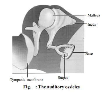
The malleus is the lateral
hammer - shaped bone. The handle is in contact with the tympanic membrane and
the head forms a movable joint with the incus.
The incus is the middle
anvil-shaped bone. Its body articulates with the malleus, the long process with
the stapes, and it is stabilized by the short process, fixed by fibrous tissue
to the posterior wall of the cavity.
The stapes is the medial
stirrup-shaped bone. Its head articulates with the incus and its base fits into
the oval window.
The three ossicles are held in position by fine ligaments.
Internal Ear
The internal ear contains the organs of hearing and balance and is
generally described in two parts, the bony labyrinth and the membranous labyrinth.
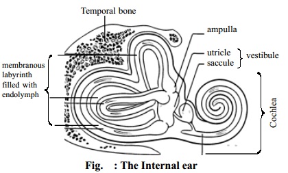
a. Bony labyrinth
This is a cavity within the temporal bone lined with periosteum. It is
larger than the membranous labyrinth of the same shape which fits into it, like
a tube within a tube. The space between the bony walls and the membranous tube
is occupied by perilymph. The
membranous labyrinth also contains fluid, the endolymph.
The bony labyrinth consists of:
1 vestibule
1 cochlea
3 semicircular canals
The vestibule is the expanded part nearest
to the middle ear. It contains the oval and round windows.
The cochlea
resembles a snail's shell. It has a broad base where it is continuous with the
vestibule and a narrow apex, and it spirals round a central bony column. The
cochlea is divided by a septum called basilar membrane.
The semicircular canals are three tubes arranged
so that one is situated in each of the three planes of space. They are
continuous with the vestibule. One end of each canal is dilated to form
ampulla. The semicircular canals are the organs of equilibrium.
b. Membranous labyrinth
The
membranous labyrinth is the same shape as its bony counterpart and is separated
from it by perilymph. It contains endolymph. It is divided into the same parts:
the vestibule which contains the utricle and saccule, the cochlea and three
semicircular canals. The utricle and saccule are oval membranous sacs.
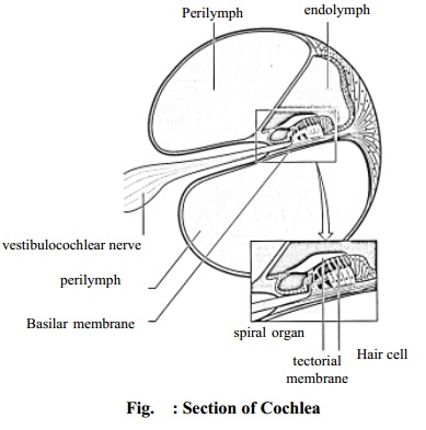
A
cross-section (Fig. 7) shows the triangular shape of the membranous cochlea.
Neuroepithelial cells and nerve fibres lie on the basilar membrane or base of
the triangle. Many of the neuroepithelial cells are long and narrow and are
arranged side by side. These cells are called hair cells and their nerve fibres
from the true organ of hearing, the organ
of Corti. The hair cells are attached to a thin membrane called tectorial membrane. The nerve fibres
combine to form the auditory part of the vestibulocochlear nerve (eighth
cranial nerve), which passes through a foramen in the temporal bone to reach
the hearing area in the temporal lobe of the cerebrum.
Related Topics