Chapter: Human Nervous System and Sensory Organs : Functional Systems
Corticospinal Tract - Motor Systems
Motor Systems
Corticospinal Tract
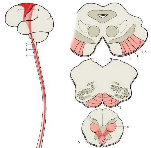
The corticospinal tract (pyramidal tract)
and the corticonuclear fibers are regarded as pathways of voluntarymovements. It is through them that the cor-tex
controls the subcortical motor centers. The cortex can have a reducing and
inhib-iting effect, but it also produces a continu-ous tonic stimulus that
promotes quick, sudden movements. The mechanical and stereotyped motions
controlled by the sub-cortical motor centers must be modified by the influence
of pyramidal impulses in order to produce specific, fine-tuned movements.
The
fibers of the corticospinal tract origi-nate in the precentral areas 4 (A1) and 6 (A2), in regions of the parietal lobe (areas 3, 1, and 2), and in
the second sensorimotor area (area 40) . Roughly
two-thirds stem from the precen-tral area, and one-third from the parietal
lobe. Only about 60% of the fibers are myeli-nated; the other 40% are
unmyelinated. The thick fibers of Betz’s giant pyramidal cells in area 4
account for only 2 – 3% of the myelinated fibers. All other fibers stem from
smaller pyramidal cells.


The
fibers of the pyramidal tract pass through the internal capsule. At the
transition to the midbrain, they ap-proach the base of the brain and together
with the corticopontine tracts form the cerebral peduncles. The corticospinal
tract fibers occupy the central part, and the fibers from the parietal cortex
occupy the most lateral position (B3).
They are followed by the corticospinal tracts for the lower limb (L, S), trunk
(T), and upper limb (C), and finally the corticonuclear fibers for the facial
region (B4). When passing through
the pons, the fiber tracts rotate so that the corticonu-clear fibers now lie
dorsally, followed by the bundles terminating in the cervical, thoracic,
lumbar, and sacral regions, respec-tively. In the medulla oblongata, the
corti-conuclear fibers terminate on the cranial nerve nuclei. Most of the
fibers.
(70 –
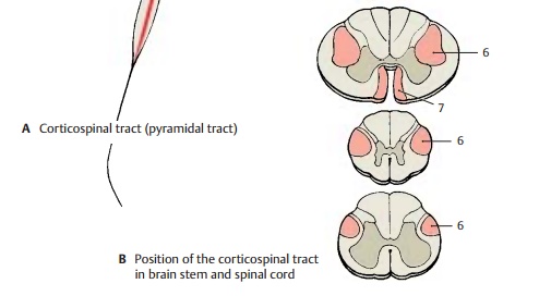
90%) cross to the opposite side in the pyramidal decussation (AB5) and form the lateral corticospinal tract (AB6).
The fibers for the upper limb cross dorsally to the fibers for the lower limb.
Within the lateral corticospinal tract, the fibers for the upper limb lie
medially, while the long fibers for the lower limb lie laterally. The uncrossed
fibers continue in the anterior
corticospinal tract (AB7) and
cross to the opposite side only at the level of their termination above the
white commissure. The anterior tract shows varia-ble degrees of development; it
may be asymmetric or even completely absent. It reaches only into the cervical
or thoracic spinal cord.
The
majority of corticospinal tract fibers terminate on interneurons in the
interme-diate zone between anterior and posterior horns. Only a small portion
reaches the motor neurons of the anterior horn, predominantly those supplying
the distal segments of the limbs, which are under the special control of the
corticospi-nal tract. Impulses from the corticospinal tract activate neurons
that innervate the flexor muscles but
inhibit neurons that innervate the extensor
muscles.
Fibers
originating from the parietal lobe terminate in the dorsal column nuclei (gracilenucleus and cuneate nucleus) and in the
sub-stantia gelatinosa of the posterior horn.They regulate the input of
sensory impulses. Hence, the corticospinal tract is not a uni-form motor
pathway but contains descending systems of different functions.
Related Topics