Chapter: Human Nervous System and Sensory Organs : Functional Systems
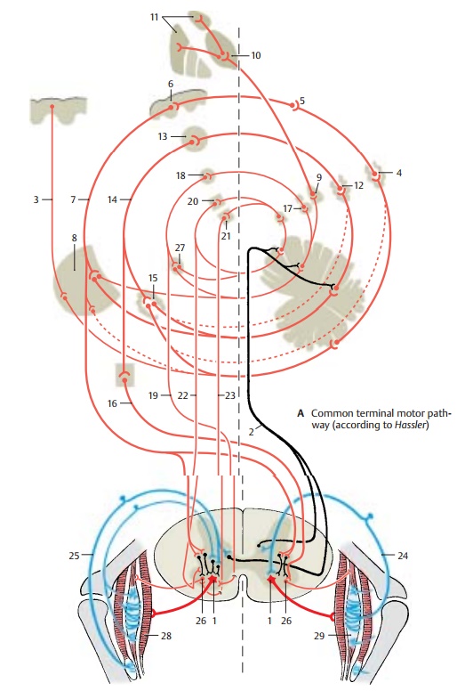
Common Terminal Motor Pathway
Common Terminal Motor Pathway
The
common terminal pathway of all centers involved in motor activity is the large
anterior horn cell (A1) and its axon (α-mo-toneuron), which innervates the voluntaryskeletal muscles. Most
of the tracts running to the anterior horn do not end directly on the anterior
horn cells but terminate on in-terneurons. The latter influence the motor
neurons either directly or act by inhibiting or activating the reflexes between
muscle receptors and motor neurons. The anterior horn is therefore not simply a
relay station as described earlier but a complex in-tegration apparatus that
regulates motor activity.
The central regions that influence motor activity via descending
pathways are inter-connected in many ways. The most impor-tant afferent
pathways stem from the cere-bellum, which receives the impulses of muscle
receptors via the spinocerebellartracts (A2) and the stimuli of the cortex
viathe corticopontine tracts (A3). The cerebellar impulses are
transmitted via the parvo-cellular part of the dentate nucleus (A4) and
the ventral lateral nucleus of thalamus
(A5) to the precentral cortex (area 4) (A6).
The corticospinal (pyramidal) tract (A7) de-scends from area 4 to the
anterior horn and gives off collaterals in the pons (A8) that re-turn
to the cerebellum. Additional cerebel-lar impulses are transmitted via the em-boliform nucleus (A9) and the centromedian nucleus of the thalamus (A10) to the striatum(A11) and via the magnocellular part of
the dentate nucleus (A12) to the red nucleus(A13). From
here fibers run in the centraltegmental
tract (A14) via the olive (A15) backto the cerebellum and in the rubroreticu-lospinal tract (A16)
to the anterior horn.Fibers from the globose
nucleus (A17) run to the interstitial nucleus of Cajal (A18) and from there in the interstitiospinal fasciculus(A19) to the anterior horn. Finally, cerebello-fugal fibers are relayed in
thevestibular nu-clei (A20) and in the reticular formation (A21)
to the vestibulospinal tract (A22) and the reticulospinal tract (A23),
respectively.

The
descending pathways can be divided into two groups according to their effect on
the muscles: one group stimulates the flexormuscles,
and another group stimulates the extensor
muscles. Thecorticospinal tractandthe
rubroreticulospinal tract activate
mainly the neurons of the flexor muscles and in-hibit the neurons of the
extensor muscles. This corresponds to the functional impor-tance of the
corticospinal tract for delicate and precise movements, especially those of
hand and finger muscles where flexor muscles play an important role. In
contrast, the fibers of the vestibulospinal
tract and the fibers from the pontine
reticular formation in-hibit the flexors and activate the extensors. They
belong to a phylogenetically old motor system that is directed against the
effect of gravity and, thus, is of special importance for body posture and
balance.
The peripheral fibers that run through the
posterior root into the anterior horn origi-nate from the muscle receptors.The
afferent fibers of the annulospiral endings (A24) ter-minate with their collaterals directly on the
α-motoneurons, while the fibers of the ten-don organs (A25) terminate on inter-neurons.Many descending pathways in-fluence
the α-neurons via the spinal reflex apparatus. They terminate on the large
α-neurons and on the small γ-neurons (A26).
Since the γ-neurons have a lower threshold of stimulation, they are stimulated
first, which results in the activation of muscle spindles. The latter send
their impulses to the α-neurons. Thus, the γ-neurons and muscle spindles have a
starter function for voluntary
movements.
A27
Accessory olive.
A28
Skeletal muscles.
A29
Muscle spindle.
Related Topics